Archive for the 'Molecules' Category

Gas-phase structure of cytosine

Alonso and coworkers have again (see this post employed laser-ablation molecular-beam Fourier-transform microwave (LA-MB-MW)spectroscopy to discern the gas phase structure of an important biological compound: cytosine.1 They identified five tautomers of cytosine 1-5. Comparison between the experimental and computational (MP2/6-311++G(d,p) microwave rotational constants and nitrogen nuclear quadrupole coupling constants led to the complete assignment of the spectra. The experimental and calculated rotational constants are listed in Table 1.

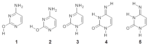
Table 1. Rotational constants (MHz) for 1-5.


1

2

3

4

5


Expt

calc

Expt

calc

Expt

calc

Expt

calc

Expt

calc

A

3951.85

3934.5

3889.46

3876.5

3871.55

3856.0

3848.18

3820.1

3861.30

3844.2

B

2008.96

1999.1

2026.32

2014.7

2024.98

2012.3

2026.31

2019.0

2011.41

1999.7

C

1332.47

1326.8

1332.87

1326.9

1330.34

1323.3

1327.99

1324.0

1323.20

1318.4

The experimental and computed relative free energies are listed in Table 2. There is both not a complete match of the relative energetic ordering of the tautomers, nor is there good agreement in their magnitude. Previous computations2 at CCSD(T)/cc-pVQZ//CCSD//cc-pVTZ are in somewhat better agreement with the gas-phase experiments.

Table 2. Relative free energies (kcal mol-1) of 1-5.


expt

MP2/
6-311++G(d,p)

CCSD(T)/cc-pVQZ//
CCSD//cc-pVTZ

1

0.0

0.0

0.0

2

0.47

0.70

0.7

3

0.11

1.19

0.2

4

0.83

3.61

0.7

5


5.22


References

(1) Alonso, J. L.; Vaquero, V.; Peña, I.; López, J. C.; Mata, S.; Caminati, W. "All Five Forms of Cytosine Revealed in the Gas Phase," Angew. Chem. Int. Ed. 2013, 52, 2331-2334, DOI: 10.1002/anie.201207744.

(2) Bazso, G.; Tarczay, G.; Fogarasi, G.; Szalay, P. G. "Tautomers of cytosine and their excited electronic states: a matrix isolation spectroscopic and quantum chemical study," Phys. Chem. Chem. Phys., 2011, 13, 6799-6807, DOI:10.1039/C0CP02354J.

InChIs

cytosine: InChI=1S/C4H5N3O/c5-3-1-2-6-4(8)7-3/h1-2H,(H3,5,6,7,8)
InChIKey=OPTASPLRGRRNAP-UHFFFAOYSA-N

MP &nucleic acids Steven Bachrach 22 Apr 2013 1 Comment

Gas—phase structure of fructose

Sugars comprise a very important class of organic compounds for a variety of reasons, including dietary needs. On the chemical side, their stereochemical variations give rise to interesting
conformational questions. While sugar structures are a well-studied dating back to Fischer, most of these studies are in the solid or solution phase, and these phases can certainly play a role in dictating conformational preferences. This is seen in the differences in conformational distribution with different solvents. Only recently has instrumentation been developed (see these posts for some earlier applications: A, B, C) that can provide structural information of sugars in the gas phase. Cocinero and co-workers describe just such an analysis of fructose.1

Using a combination of laser ablation Fourier transform microwave spectroscopy and quantum chemical computations, they have examined the gas-phase structure of this ketose. There are
quite a number of important conformational and configurational isomers to consider, as shown in Scheme 1. Fructose can exist in a pyranose form (6-member ring) with the anomeric carbon being α or β. An alternative cyclic form is the 5-member ring furanose form, which again has the two options at the anomeric position. Both the 5- and 6-member rings can ring flip, giving rise to 4 pyranose and 4 furanose forms. Of course there is also the
acyclic form.

Scheme 1. Major Frucotse isomers

α-pyranose

β-pyranose

α-furanose

β-furanose

Open chain

The observed microwave spectrum is quite simple, showing evidence of only a single isomer. In comparing the microwave rotational constants and the quartic centrifugal distortion constants with those obtained from MP2 and M06-2x computations, it is clear that the only observed isomer is the β-pyranose isomer in its 2C5 conformation.

Both MP2 and M06-2x (with a variety of TZ basis sets) predict this isomer to be the lowest energy form by about 2.5 kcal mol-1. This structure is shown in Figure 1. Interestingly, B3LYP predicts the open chain configuration as the most stable isomer, with the β-pyranose isomer about 0.5 kcal mol-1 higher in energy. The authors strongly caution against using B3LYP for any sugars.

Figure 1. MP2/maug-cc-pVTZ optimized structure of β-fructopyranose.

This most stable furanose isomer displays five intramolecular hydrogen bonds that account for its stability over all other possibilities. However, the pyranose form of fructose is very rare in nature, and the Protein Data Bank has only four examples. The furanose form is by far the more commonly found isomer (as in sucrose). Clearly, hydrogen bonding to solvent and other solvent interactions alter the conformational distribution.

References

(1) Cocinero, E. J.; Lesarri, A.; Ecija, P.; Cimas, A.; Davis, B. G.; Basterretxea, F. J.; Fernandez, J. A.; Castano, F. "Free Fructose is Conformationally Locked," J. Am. Chem. Soc. 2013, 135, 2845-2852, DOI: 10.1021/ja312393m.

InChIs:

β-fructopyranose:
InChI=1S/C5H10O6/c6-2-1-11-5(9,10)4(8)3(2)7/h2-4,6-10H,1H2
InChIKey=FFDHYUUPNCCTDA-UHFFFAOYSA-N

sugars Steven Bachrach 14 Mar 2013 2 Comments

Monosaccharides benchmark

A comprehensive evaluation of how different computational methods perform in predicting the energies of monosaccharides comes to some very interesting conclusions. Sameera and Pantazis1 have examined the eight different aldohexoses (allose, alltrose, glucose, mannose, gulose, idose, galactose and talose), specifically looking at different rotomers of the hydroxymethyl group, α- vs. β-anomers, pyranose vs. furanose isomers, ring conformations (1C4 vs skew boat forms), and ring vs. open chain isomers. In total, 58 different structures were examined. The benchmark computations are CCSD(T)/CBS single point energies using the SCS-MP2/def2-TZVPP optimized geometries. The RMS deviation from these benchmark energies for some of the many different methods examined are listed in Table 1.

Table 1. Average RMS errors (kJ mol-1) of the 58 different monosaccharide structures for
different computational methods.

method

average RMS error

LPNO-CEPA

0.71

MP2

1.27

SCS-MP2

1.55

mPW2PLYP-D

2.02

M06-2x

2.03

PBE0

3.62

TPSS

4.78

B3LYP-D

4.79

B3LYP

5.06

HF

6.69

B97D

7.66

Perhaps the most interesting take-home message is that CEPA, MP2, the double hybrid methods and M06-2x all do a very good job at evaluating the energies of the carbohydrates. Given the significant computational advantage of M06-2x over these other methods, this seems to be the functional of choice! The poorer performance of the DFT methods over the ab initio methods is primarily in the relative energies of the open-chain isomers, where errors can be on the order of 10-20 kJ mol-1 with most of the functionals; even the best overall methods (M06-2x and the double hybrids) have errors in the relative energies of the open-chain isomers of 7 kJ mol-1. This might be an area of further functional development to probe better treatment of the open-chain aldehydes vs. the ring hemiacetals.

References

(1) Sameera, W. M. C.; Pantazis, D. A. "A Hierarchy of Methods for the Energetically Accurate Modeling of Isomerism in Monosaccharides," J. Chem. Theory Comput. 2012, 8, 2630-2645, DOI:10.1021/ct3002305

DFT &sugars Steven Bachrach 28 Nov 2012 No Comments

Amino acid-catalyzed aldol and Michael reactions

Here are a couple of articles describing computational approaches to catalytic enantioselective reactions using variations upon the classic proline-catalyzed aldol reaction of List and Barbas1 that started the whole parade. I have discussed the major computational papers on that system in my book (Chapter 5.3).

Yang and Wong2 investigated the proline-catalyzed nitro-Michael reaction, looking at four examples, two with aldehydes and two with ketones (Reactions 1-4).

Reaction 1

Reaction 2

Reaction 3

Reaction 4

These four reactions were examined atMP2/311+G**//M06-2x/6-31G**, and PCM was also applied. The key element of this study is that they examined two different types of transition states: (a) based on the Houk-List model involving a hydrogen bond and (b) an electrostatic based model with no hydrogen bond. These are sketched in Scheme 1. For each of the reactions 1-4 there are 8 located transition states differing in the orientation of the attack on to the syn or anti enamine.

Scheme 1. TS models


Model A


Model B

The two lowest energy TS are shown in Figure 1. TS1-β1-RS is the lowest TS and it leads to the major enantiomer. The second lowest TS, TS1-β3-SR, lies 2.9 kJ mol-1 above the other TS, and it leads to the minor enantiomer. This lowest TS is of the Houk-List type (Model A) while the other TS is of the Model B type. The enthalpies of activation suggest an ee of 54%, in reasonable
agreement with experiment.

TS1-β1-RS

TS1-β1-RS

Figure 1. M06-2x/6-31G** optimized geometries of TS1-β1-RS and TS1-β1-RS.

The computations of the other three reactions are equally good in terms of agreement with experiment, and importantly the computations indicate the reversal of stereoselection between the aldehydes and the ketones. These computations clearly implicate both the Houk-List and the non-hydrogen bonding TSs in the catalyzed Michael additions.

Houk in collaboration with Scheffler and Mahrwald investigate the use of histidine as a catalyst for the asymmetricaldol reaction.3 Examples of the histidine-catalyzed aldol are shown in Reactions 5-7.

Reaction 5

Reaction 6

Reaction 7

The interesting twist here is whether the imidazole can also be involved in hydrogen bonding to the acceptor carbonyl group, serving the purpose of the carboxylic acid group in the Houk-List TS when proline is the catalyst (Model A). The transition states for these and a few other reactions were computed at M06-2x/6-31+G(d,p) including with the SMD continuum solvent model for water. The two lowest energy TSs for the reaction of isobutyraldehde and formaldehyde are shown in Figure 2; TS1 has the carboxylic acid group as the hydrogen donor while the imidazole is the donor in TS2. Of note is that these two TS are isoenergetic, indicating that both modes of stabilization are at play with histidine as the catalyst.

TS1

TS2

Figure 2. M06-2x/6-31+G(d,p) geometries of the two lowest energy TSs for the reaction of isobutyraldehde and formaldehyde catalyzed by histidine.

The possible TSs for Reactions 5-7 were also located. For example, with Reaction 5, the lowest energy TS involves the imidazole as the hydrogen donor and it leads to the major product. The lowest energy TS that leads to the minor product involves the carboxylic acid as the donor. The computed ee’s for Reactions 5-7 are in very good, if not excellent, agreement with the experimental values. The study should spur further activity in which one might tune the stereoselectivity by using catalysts with multiple binding opportunities.

References

(1) List, B.; Lerner, R. A.; Barbas, C. F., III; "Proline-Catalyzed Direct Asymmetric Aldol Reactions," J. Am. Chem. Soc., 2000, 122, 2395-2396, DOI: 10.1021/ja994280y.

(2) Yang, H.; Wong, M. W. "(S)-Proline-catalyzed nitro-Michael reactions: towards a better understanding of the catalytic mechanism and enantioselectivity," Org. Biomol. Chem.,
2012, 10, 3229-3235, DOI: 10.1039/C2OB06993H

(3) Lam, Y.-h.; Houk, K. N.; Scheffler, U.; Mahrwald, R. "Stereoselectivities of Histidine-Catalyzed Asymmetric Aldol Additions and Contrasts with Proline Catalysis: A Quantum Mechanical Analysis," J. Am. Chem. Soc. 2012, 134, 6286-6295, DOI: 10.1021/ja2118392

aldol &amino acids &Houk &Michael addition &stereoinduction Steven Bachrach 15 May 2012 1 Comment

Fluoresence of encapsulated stilbene

Petsalakis and Rebek have explored the fluorescence of stilbene inside a couple of different kinds of capsules.1 trans-Stilbene exhibits weak fluorescence in solution, but when placed inside a small capsule, the fluorescence disappears almost entirely, while in a large capsule, the fluorescence returns to normal. They examined stilbene inside two different capsules using a variety of DFT and ONIOM techniques.

The optimized geometries of trans- and cis-stilbene optimized at CAM-B3LYP/6-31G(d,p) are displayed in Figure 1. As expected, the trans conformer is planar and the cis conformer is twisted to avoid clashes between the phenyl rings. The optimized structure of the 1.1 capsule is also shown in Figure 1. All of these structures are fairly insensitive to computational method. (They have also looked at an even larger capsule, but I have omitted displaying its structure here.)

(a)

(b)

(c)

(d)

Figure 1. CAM-B3LYP/6-31G(d,p) optimized structures of (a) trans-stilbene, (b) cis-stilbene , (c) the 1.1 capsule, and (d) trans-stilbene inside the 1.1 capsule.

The structure of trans- stilbene inside the 1.1 capsule is shown in Figure 1. Of particular note is that the stilbene is no longer planar. (This twisting is perhaps better observed by an end-on view, which the reader can obtain by clicking on the picture and then manipulating the full 3-D structure using the Jmol applet.) The different computational methods give slightly different encapsulated structures, and vary a bit in their binding energies, but the twisting of the stilbene is reproduced by each method. Though not shown here, trans-stilbene in the larger capsule is again a nearly planar structure.

The structure of the S1 state of trans-stilbene in the large capsule is the same as for free trans-stilbene. However, the geometry of the S1 state in the smaller 1.1 capsule is twisted and corresponds to the conical intersection geometry.

The absorption spectra and the emission spectra were computed for the free and encapsulated structures. The absorption and emission spectra for free stilbene and stilbene in the larger capsule are nearly identical, corresponding to the experimental observation of similar fluorescence behavior. The absorption spectra of stilbene in the 1.1 capsule has a small blue shift of 8 nm due to the twisted geometry. But the major result is that the S1 state of stilbene inside the 1.1 capsule distorts to the conical intersection, allowing for radiationless return to the ground state. This means that there would be no fluorescence, and that is exactly what is observed.

References

(1) Tzeli, D.; Theodorakopoulos, G.; Petsalakis, I. D.; Ajami, D.; Rebek, J., "Conformations and Fluorescence of Encapsulated Stilbene," J. Am. Chem. Soc., 2012, 134, 4346-4354, DOI: 10.1021/ja211164b

InChIs

trans-stilbene: InChI=1/C14H12/c1-3-7-13(8-4-1)11-12-14-9-5-2-6-10-14/h1-12H/b12-11+
InChIKey=PJANXHGTPQOBST-VAWYXSNFBV

cis-stilbene: InChI=1/C14H12/c1-3-7-13(8-4-1)11-12-14-9-5-2-6-10-14/h1-12H/b12-11-
InChIKey=PJANXHGTPQOBST-QXMHVHEDBW

stilbene Steven Bachrach 10 Apr 2012 No Comments

Nanohoop of linked napthlylene groups

Itami continues to design novel macrocycles containing aromatic rings (see this post). This latest paper reports the synthesis of the first nanohoops containing naphthylenes, namely [9]cyclo-1,4-naphthylene 1.1 Since the macrocycle contains an odd number of naphthylene units, the lowest energy conformation is of C2 symmetry with one of the naphthylene rings in the plane of the macrocycle. (See Figure 1 for the B3LYP/6-31G(d) optimized structure). This conformation gives rise to 27 peaks in the proton NMR, and while the value of the computed chemical shifts differ from the experimental ones by about 0.5 to 1 ppm, their relative ordering is in very nice agreement.

1

2

Figure 1. B3LYP/6-31G(d) optimized geometries of 1 and the racemization transition state 2.

Itami also notes that 1 is chiral and computed the barrier for racemization of 19.9 kcal mol-1¸ through the transition state 2, also shown in Figure 1. This racemization process is compared with the racemization of 1,1’-binaphthyl.

References

(1) Yagi, A.; Segawa, Y.; Itami, K., "Synthesis and Properties of [9]Cyclo-1,4-naphthylene: A π-Extended Carbon Nanoring," J. Am. Chem. Soc. 2012, 134, 2962-2965, DOI: 10.1021/ja300001g

InChIs

1: InChI=1/C90H54/c1-2-20-56-55(19-1)73-37-38-74(56)76-41-42-78(60-24-6-5-23-59(60)76)80-45-46-82(64-28-10-9-27-63(64)80)84-49-50-86(68-32-14-13-31-67(68)84)88-53-54-90(72-36-18-17-35-71(72)88)89-52-51-87(69-33-15-16-34-70(69)89)85-48-47-83(65-29-11-12-30-66(65)85)81-44-43-79(61-25-7-8-26-2(61)81)77-40-39-75(73)57-21-3-4-22-58(57)77/h1-54H/b75-73-,76-74-,79-77-,80-78-,83-81-,84-82-,87-85-,88-86-,90-89-
InChIKey=WUFIQFYLEOBLMY-YNQZQJAJBX

nanohoops Steven Bachrach 27 Mar 2012 No Comments

Ethynyl-substituted Cyclobutadiene

Cyclobutadiene is the prototypical antiaromatic compound. McMahon has examined the
effect of ethynyl substitution on this ring, with a long term eye towards the possibility of these types of species being involved in the synthesis of fullerenes.1

All of the possible ethynyl-substituted cyclobutadiene species (1-7) were optimized at B3LYP/6-31G(d) and CCSD/cc-pVDZ in their singlet and triplet states.

The structures of singlet and triplet 7 are shown in Figure 1. The geometries provided by the two different methods are quite similar. They show a rectangular form for the singlets and a delocalized, nearly square ring for the triplets.

7singlet

7triplet

Figure 1. CCSD/cc-pVDZ optimized structures of singlet and triplet 7.

The computed singlet-triplet gap decreases with each ethynyl substituent. B3LYP, which overestimates the stability of triplets, predicts that 6 and 7 will be ground state triplets, while CCSD predicts a singlet ground state for all 7 species, with the gap decreasing steadily from 11.5 to 8.2 kcal mol-1, a value that is also probably underestimated.

This change in the singlet-triplet gap reflects a stronger stabilizing effect of each ethynyl group to the cycnobutadiene ring for the triplet than for the singlet state. This is seen in the homodesmotic stabilization energies.

Lastly, NICS(1)zz values are positive for all of the singlets and negative for the triplets. The positive values for the singlets reflect their antiaromatic character, also seen in the alternant bond distances around the ring. The NICS values of the singlets decrease with increasing substitution. The negative NICS values of the triplets reflects aromatic character, as seen in the non-alternant distances around the ring. Interestingly, the triplet NICS values decrease with increasing ethynyl substitution, suggesting decreased aromaticity, even though the homodesmotic reactions suggest increasing stabilization with substitution.

References

(1) Esselman, B. J.; McMahon, R. J., "Effects of Ethynyl Substitution on Cyclobutadiene," J. Phys. Chem. A 2012, 116, 483-490, DOI: 10.1021/jp206478q

InChIs

1: InChI=1/C4H4/c1-2-4-3-1/h1-4H
InChIKey=HWEQKSVYKBUIIK-UHFFFAOYAI

2: InChI=1/C6H4/c1-2-6-4-3-5-6/h1,3-5H
InChIKey=XFHXCHFBSJDBGT-UHFFFAOYAG

3: InChI=1/C8H4/c1-3-7-5-6-8(7)4-2/h1-2,5-6H
InChIKey=YSSBCMLQKUIAEP-UHFFFAOYAI

4: InChI=1/C8H4/c1-3-7-5-8(4-2)6-7/h1-2,5-6H
InChIKey=IRAQOGPKMAXTQY-UHFFFAOYAN

5: InChI=1/C8H4/c1-3-7-5-6-8(7)4-2/h1-2,5-6H
InChIKey=YSSBCMLQKUIAEP-UHFFFAOYAI

6: InChI=1/C10H4/c1-4-8-7-9(5-2)10(8)6-3/h1-3,7H
InChIKey=BVCIPPXDFXWTJR-UHFFFAOYAQ

7: InChI=1/C12H4/c1-5-9-10(6-2)12(8-4)11(9)7-3/h1-4H
InChIKey=HEUYILYXFVQGOW-UHFFFAOYAO

cyclobutadiene Steven Bachrach 20 Mar 2012 No Comments

Singlet-triplet carbene gap and remote subtituents

Can a remote substituent affect the singlet-triplet spin state of a carbene? Somewhat surprisingly, the answer is yes. Sheridan has synthesized and characterized the meta and para methoxy-substituted phenyltrifluoromethyl)carbenes 1 and 2.1 The UV-Vis spectrum of 1 is consistent with a triplet as its EPR and reactivity with oxygen. However, the para isomer 2 gave no EPR signal and failed to react with oxygen or hydrogen, suggestive of a singlet.

The conformations of 1 and 2 were optimized at B3LYP/6-31+G(d,p) and the lowest energy
singlet and triplet conformers are shown in Figure 1. The experimental spectral features of 1 match up best with the computed features of the triplet, and the same is true for the singlet of 2.

1singlet

1triplet

2singlet

2triplet

Figure 1. B3LYP/6-31+G(d,p) optimized geometries of 1 and 2.

The triplet of 1 is estimated to be about 4 kcal mol-1 below that of the singlet – larger than the general overestimation of the stability of triplets that beleaguer B3LYP. For 2, B3LYP predicts a singlet ground state.

The isodesmic reactions 1 and 2 help understand the strong substituent effect. For 1, the meta substituent destabilizes both the singlet and triplet by a small amount. For 2, the para methoxy group stabilizes the triplet slightly, but stabilizes the singlet by a large amount. This stabilization is likely the result of the contribution of a second resonance structure 2b. A large rotational barrier for both the methyl methyl and the trifluoromethyl groups supports the participation of 2b.


ΔEsinglet = -0.8 kcal mol-1
ΔEtriplet = -0.6 kcal mol-1

Rxn 1


ΔEsinglet = -5.8 kcal mol-1
ΔEtriplet = -1.1 kcal mol-1

Rxn 2

References

(1) Song, M.-G.; Sheridan, R. S., "Regiochemical Substituent Switching of Spin States in
Aryl(trifluoromethyl)carbenes," J. Am. Chem. Soc. 2011, 133, 19688-19690, DOI: 10.1021/ja209613u

InChIs

1: InChI=1/C9H7F3O/c1-13-8-4-2-3-7(5-8)6-9(10,11)12/h2-5H,1H3
InChIKey=CAHMVAJXXQQHDW-UHFFFAOYAA

2: InChI=1/C9H7F3O/c1-13-8-4-2-7(3-5-8)6-9(10,11)12/h2-5H,1H3
InChIKey

carbenes Steven Bachrach 14 Feb 2012 4 Comments

tri-p-quinodimethane

Tsuji and Nakamura have prepared the tri-p­-quinodimethane 1.1 Quinodimethanes are of interest because of their possible diradical character. This new example is most interesting. It is stable as a solid in air and ambient light for 6 months, or 2 months in solution. Its ESR shows fine structure, with a spin-spin distance estimated to be 14.6 Å, very close to the distance between the terminal carbons. The ground state is a singlet, with the triplet lying 2.12 kcal mol-1 higher in energy.


Ar = 4-octylphenyl

UB3LYP/6-31G** computations (lacking the aryl and phenyl sidechains) indicate a ground state singlet (with sizable spin contamination) and a gap to the triplet of 1.83 kcal mol-1. The computed geometry is shown in Figure 1.

1

Figure 1. UB3LYP/6-31G** optimized geometry of 1.

The analog having just two quinodimethane units showed no ESR signal and the computed singlet-triplet energy gap is 5.68 kcal mol-1.

It would have been interesting to have computed the NICS values for the 6-member rings – as a measure of aromatic vs. non aromatic character to further support the participation of the biradical resonance structure contribution to 1.

References

(1) Zhu, X.; Tsuji, H.; Nakabayashi, K.; Ohkoshi, S.-i.; Nakamura, E., "Air- and Heat-Stable Planar Tri-p-quinodimethane with Distinct Biradical Characteristics," J. Am. Chem. Soc., 2011, 133, 16342-16345, DOI: 10.1021/ja206060n

InChI

1: InChI=1/C112H112N4/c1-5-9-13-17-21-29-41-81-53-63-93(64-54-81)111(94-65-55-82(56-66-94)42-30-22-18-14-10-6-2)101-73-85(87(77-113)78-114)61-71-97(101)105-107(111)99-75-104-100(76-103(99)109(105,89-45-33-25-34-46-89)90-47-35-26-36-48-90)108-106(110(104,91-49-37-27-38-50-91)92-51-39-28-40-52-92)98-72-62-86(88(79-115)80-116)74-102(98)112(108,95-67-57-83(58-68-95)43-31-23-19-15-11-7-3)96-69-59-84(60-70-96)44-32-24-20-16-12-8-4/h25-28,33-40,45-76H,5-24,29-32,41-44H2,1-4H3
InChIKey=IDOIPCRGROEZHG-UHFFFAOYAD

1 (lacking aryl side chains): InChI=1/C32H16N4/c33-13-23(14-34)17-1-3-25-19(5-17)9-31-27-8-22-12-30-26-4-2-18(24(15-35)16-36)6-20(26)10-32(30)28(22)7-21(27)11-29(25)31/h1-8H,9-12H2
InChIKey=JRLKUOJPPWAWTD-UHFFFAOYAN

diradicals Steven Bachrach 28 Nov 2011 1 Comment

Structure of 1-aminocyclopropylcarboxylic acid

There are three generic conformations of α-amino acids in the gas phase: A-C. These are stabilized by intramolecular hydrogen bonds. While computations suggest that all three are close in energy, the very detailed laser ablation- molecular beam-Fourier transform microwave (LA-MB-FTMW) experiments of the Alonso group (mentioned in these previous posts: guanine, cysteine, ephedrine) have identified only the first two conformations. Cooling of the structures in the jet expansion appears to be the reason for the loss of the (slightly) higher energy conformer C.

Alonso now reports on the structure of 1-aminocyclopropanecarboxylic acid 1.1 The three MP2/6-311+G(d,p) optimized conformation are shown in Figure 1. The interaction between the cyclopropyl orbitals and the carbonyl π-bond suggests that only two structures (where the carbonyl bisects thecyclopropyl plane) will exist and that rotation between them may require passage through a prohibitively high barrier. In fact, computations suggest a barrier of 2000 cm-1 (5.7 kcal mol-1). This is much larger than the typical rotation barrier of the amino acids that interconvert A with C, which are about 400 cm-1 (1 kcal mol-1).

1A

1B

1C

After careful examination of the microwave spectrum, all three conformations 1A-C were identified by comparing the experimental value of the rotational constants, and 14N nuclearquadrupole coupling constants with the computed values. Really excellent agreement is found, including in the ratio of the relative amounts of the three isomers. Once again, we have an exquisite example of the importance of computations and experiments being used in conjunction to solve interesting chemical problems.

References

(1) Jimenez, A. I.; Vaquero, V.; Cabezas, C.; Lopez, J. C.; Cativiela, C.; Alonso, J. L., "The Singular Gas-Phase Structure of 1-Aminocyclopropanecarboxylic Acid (Ac3c)," J. Am. Chem. Soc., 2011, 133, 10621-10628, DOI: 10.1021/ja2033603

InChIs

1: InChI=1/C4H7NO2/c5-4(1-2-4)3(6)7/h1-2,5H2,(H,6,7)/f/h6H
InChIKey=PAJPWUMXBYXFCZ-BRMMOCHJCR

amino acids Steven Bachrach 04 Oct 2011 No Comments

« Previous PageNext Page »