Archive for the 'NMR' Category

dJ-DP4 and iJ-DP4: including coupling constants

I have written quite a number of posts on using quantum mechanics computations to predict NMR spectra that can aid in identifying chemical structure. Perhaps the most robust technique is Goodman’s DP4 method (post), which has seen some recent revisions (updated DP4, DP4+). I have also posted on the use of computed coupling constants (posts).

Grimblat, Gavín, Daranas and Sarotti have now combined these two approaches, using computed 1H and 13C chemical shifts and 3JHH coupling constants with the DP4 framework to predict chemical structure.1

They describe two different approaches to incorporate coupling constants:

  • dJ-DP4 (direct method) incorporates the coupling constants into a new probability function, using the coupling constants in an analogous way as chemical shifts. This requires explicit computation of all chemical shifts and 3JHH coupling constants for all low-energy conformations.
  • iJ-DP4 (indirect method) uses the experimental coupling constants to set conformational constraints thereby reducing the number of total conformations that need be sampled. Thus, large values of the coupling constant (3JHH > 8 Hz) selects conformations with coplanar hydrogens, while small values (3JHH < 4 Hz) selects conformations with perpendicular hydrogens. Other values are ignored. Typically, only one or two coupling constants are used to select the viable conformations.

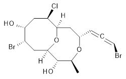
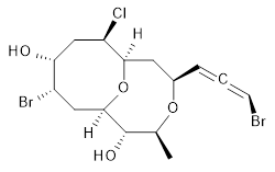
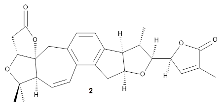
The authors test these two variants on 69 molecules. The original DP4 method predicted the correct stereoisomer for 75% of the examples, while dJ-DP4 correct identifies 96% of the cases. As a test of the indirect method, they examined marilzabicycloallenes A and B (1 and 2). DP4 predicts the correct stereoisomer with only 3.1% (1) or <0.1% (2) probability. dJ-DP4 predicts the correct isomer for 1 with 99.9% probability and 97.6% probability for 2. The advantage of iJ-DP4 is that using one coupling constant reduces the number of conformations that must be computed by 84%, yet maintains a probability of getting the correct assignment at 99.2% or better. Using two coupling constants to constrain conformations means that only 7% of all of the conformations need to be samples, and the predictive power is maintained.

1

2

Both of these new methods clearly deserve further application.

References

1. Grimblat, N.; Gavín, J. A.; Hernández Daranas, A.; Sarotti, A. M., “Combining the Power of J Coupling and DP4 Analysis on Stereochemical Assignments: The J-DP4 Methods.” Org. Letters 2019, 21, 4003-4007, DOI: 10.1021/acs.orglett.9b01193.

InChIs

1: InChI=1S/C15H21Br2ClO4/c1-8-15(20)14-6-10(17)12(19)7-11(18)13(22-14)5-9(21-8)3-2-4-16/h3-4,8-15,19-20H,5-7H2,1H3/t2-,8-,9+,10-,11+,12+,13+,14+,15-/m0/s1
InChIKey=APNVVMOUATXTFG-NTSAAJDMSA-N

2: InChI=1S/C15H21Br2ClO4/c1-8-15(20)14-6-10(17)12(19)7-11(18)13(22-14)5-9(21-8)3-2-4-16/h3-4,8-15,19-20H,5-7H2,1H3/t2-,8-,9-,10-,11+,12+,13+,14+,15-/m0/s1
InChIKey=APNVVMOUATXTFG-SSBNIETDSA-N
NMR Steven Bachrach 26 Jun 2019 Comments Off on dJ-DP4 and iJ-DP4: including coupling constants

New Procedure for computing NMR spectra with spin-spin coupling

Computed NMR spectra have become a very useful tool in identifying chemical structures. I have blogged on this multiple times. A recent trend has been the development of computational procedures that lead to computed spectra (again, see that above link). Now, Grimme, Neese and coworkers have offered their approach to computed NMR spectra, including spin-spin splitting.1

Their procedure involves four distinct steps.

  1. Generation of the conformer and rotamer space. This is a critical distinctive element of their method in that they take a number of different tacks for sampling conformational space to insure that they have identified all low-energy structures. This involves a combination of normal mode following, genetic structure crossing (based on genetic algorithms for optimization), and molecular dynamics. Making this all work is their choice of using the computational efficient GFN-xTB2 quantum mechanical method.
  2. The low-energy structures are then subjected to re-optimization at PBEh-3c and then single-point energies obtained at DSD-BLYP-D3/def2-TZVPP including treatment of solvation by COSMO-RS. The low-energy structures that contribute 4% or more of the Boltzmann-weighted population are then carried forward.
  3. Chemical shifts and spin-spin coupling constants are then computed with the PBE0 method and the pcS and pcJ basis sets developed by Jensen for computing NMR shifts.3
  4. Lastly, the chemical shifts and coupling constants are averaged and the spin Hamiltonian is solved.

The paper provides a number of examples of the application of the methodology, all with quite good success. The computer codes to run this method are available for academic use from xtb@thch.uni-bonn.de.

References

1) Grimme, S.; Bannwarth, C.; Dohm, S.; Hansen, A.; Pisarek, J.; Pracht, P.; Seibert, J.; Neese, F., "Fully Automated Quantum-Chemistry-Based Computation of Spin–Spin-Coupled Nuclear Magnetic Resonance Spectra." Angew. Chem. Int. Ed. 2017, 56, 14763-14769, DOI: 10.1002/anie.201708266.

2) Grimme, S.; Bannwarth, C.; Shushkov, P., "A Robust and Accurate Tight-Binding Quantum Chemical Method for Structures, Vibrational Frequencies, and Noncovalent Interactions of Large Molecular Systems Parametrized for All spd-Block Elements (Z = 1–86)." J. Chem. Theory Comput. 2017, 13, 1989-2009, DOI: 10.1021/acs.jctc.7b00118.

3) Jensen, F., "Basis Set Convergence of Nuclear Magnetic Shielding Constants Calculated by Density Functional Methods." J. Chem. Theory Comput. 2008, 4, 719-727, DOI: 10.1021/ct800013z.

Grimme &NMR Steven Bachrach 05 Feb 2018 No Comments

More applications of computed NMR spectra

In this post I cover two papers discussing application of computed NMR chemical shifts to structure identification and (yet) another review of computational techniques towards NMR structure prediction.

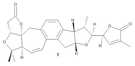
Grimblat, Kaufman, and Sarotti1 take up the structure of rubriflordilactone B 1, which was isolated from Schisandra rubriflora. The compound was then synthesized and its x-ray structure reported, however its NMR did not match with the natural extract. It was suggested that there were actually two compounds in the extract, the minor one was less soluble and is the crystallized 1, and a second compound responsible for the NMR signal.

The authors looked at all stereoisomers of this molecule keeping the three left-most rings intact. The low energy rotamers of these 32 stereoisomers were then optimized at B3LYP/6-31G* and the chemical shifts computed at PCM(pyridine)/mPW1PW91/6-31+G**. To benchmark the method, DP4+ was used to identify which stereoisomer best matches with the observed NMR of authentic 1; the top fit (92.6% probability) was the correct structure.

The 32 stereoisomers were then tested against the experimental NMR of the natural extract. DP4+ with just the proton shifts suggested structure 2 (99.8% probability); however, the 13C chemical shifts predicted a different structure. Re-examination of the reported chemical shifts identifies some mis-assigned signals, which led to a higher C-DP4+ prediction. When all 128 stereoisomers were tested, structure 2 had the highest DP4+ prediction (99.5%), but the C-DP4+ prediction remained problematic (10.8%). Analyzing the geometries of all reasonable alternative for agreement with the NOESY spectrum confirmed 2. These results underscore the importance of using all data sources.

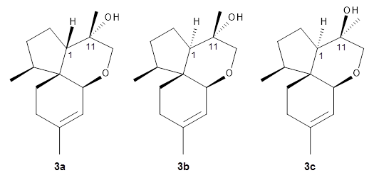
Reddy and Kutateladze point out the importance of using coupling constants along with chemical shifts in structure identification.2 They examined cordycepol A 3, obtained from Cordyceps ophioglossoides. They noted that the computed chemical shifts and coupling constants of originally proposed structure 3a differed dramatically from the experimental values.

They first proposed that the compound has structure 3b. The computed coupling constants using their relativistic force field.3 The experimental coupling constants for the proton H1 are 13.4 and 7.1 Hz. The computed values for 3a are 8.9 and 1.6 Hz, and this structure is clearly incorrect. The coupling constants are improved with 3b, but the 13C chemical shifts are in poor agreement with experiment. So, they proposed structure 3c, the epimer at both C1 and C11 of the original structure.

They optimized four conformations of 3c at B3LYP/6-31G(d) and obtained Boltzmann-weighted chemical shifts at mPW1PW91/6-311+G(d,p). The RMS deviation of the computed 13C chemical shifts relative to the experiment is only 1.54 ppm, and more importantly, the computed coupling constants of 13.54 and 6.90 Hz are in excellent agreement with the experiment values.

Lastly, Grimblat and Sarotti present a review of a number of methods for using computed NMR chemical shifts towards structure prediction.4 These methods include CP3, DP4, DP4+ (all of which I have posted on in the past) and an artificial neural network approach of their own design. They discuss a number of interesting cases where each of these methods has been crucial in identifying the correct chemical structure.

References

1. Grimblat, N.; Kaufman, T. S.; Sarotti, A. M., "Computational Chemistry Driven Solution to Rubriflordilactone B." Org. Letters 2016, 18, 6420-6423, DOI: 10.1021/acs.orglett.6b03318.

2. Reddy, D. S.; Kutateladze, A. G., "Structure Revision of an Acorane Sesquiterpene Cordycepol A." Org. Letters 2016, 18, 4860-4863, DOI: 10.1021/acs.orglett.6b02341.

3. (a) Kutateladze, A. G.; Mukhina, O. A., "Minimalist Relativistic Force Field: Prediction of Proton–Proton Coupling Constants in 1H NMR Spectra Is Perfected with NBO Hybridization Parameters." J. Org. Chem. 2015, 80, 5218-5225, DOI: 10.1021/acs.joc.5b00619; (b) Kutateladze, A. G.; Mukhina, O. A., "Relativistic Force Field: Parametrization of 13C–1H Nuclear Spin–Spin Coupling Constants." J. Org. Chem. 2015, 80, 10838-10848, DOI: 10.1021/acs.joc.5b02001.

4. Grimblat, N.; Sarotti, A. M., "Computational Chemistry to the Rescue: Modern Toolboxes for the Assignment of Complex Molecules by GIAO NMR Calculations." Chem. Eur. J. 2016, 22, 12246-12261, DOI: h10.1002/chem.201601150.

InChIs

1: InChI=1S/C28H30O6/c1-13-9-20(32-26(13)30)25-14(2)24-17-6-5-15-12-28-21(8-7-16(15)18(17)10-19(24)31-25)27(3,4)33-22(28)11-23(29)34-28/h5-9,14,19-22,24-25H,10-12H2,1-4H3/t14-,19+,20-,21-,22+,24-,25-,28+/m0/s1
InChIKey=JGSLSHOXBXVVTQ-NEUKEVNNSA-N

2: InChI=1S/C28H30O6/c1-13-9-20(32-26(13)30)25-14(2)24-17-6-5-15-12-28-21(8-7-16(15)18(17)10-19(24)31-25)27(3,4)33-22(28)11-23(29)34-28/h5-9,14,19-22,24-25H,10-12H2,1-4H3/t14-,19-,20-,21-,22+,24+,25-,28+/m0/s1
InChIKey=JGSLSHOXBXVVTQ-WQIRXNRDSA-N

3c: InChI=1S/C16H28O2/c1-6-11(2)9-14-16(5)12(3)7-8-13(16)15(4,17)10-18-14/h9,12-14,17H,6-8,10H2,1-5H3/b11-9-/t12-,13-,14-,15-,16+/m0/s1
InChIKey=WPQIVUHVYBQTBG-AWEVENECSA-N

NMR Steven Bachrach 04 Oct 2017 No Comments

Another procedure for computing NMR chemical shifts

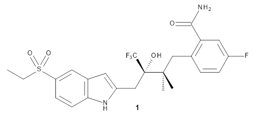
Here’s another take on automating a procedure for using computer 13C chemical shifts to assess chemical structure.1 (Have a look at these previous posts for some alternative methods and applications.) The approach here is to benchmark a few computational methods against a conformationally flexible drug-like molecule, in this case 1. A variety of conformations were optimized using the different computational methods, and 13C chemical shifts evaluated from a Boltzmann-weighted distribution. While the best agreement with the experimental chemical shifts (based on the root-mean-squared deviation) is with ωB97XD/cc-pVDZ, the authors opt for B3LYP/cc-pVDZ for its computational efficiency with only slightly poorer performance. (It should be note that WC04/cc-pVDZ, a functional designed for computing 13 chemical shifts,2 is almost as good as ωB97XD/cc-pVDZ. Also, not mentioned in the article is the dramatically poorer performance of the pcS-2 basis set, despite the fact that it was parametrized3 for NMR computation!)

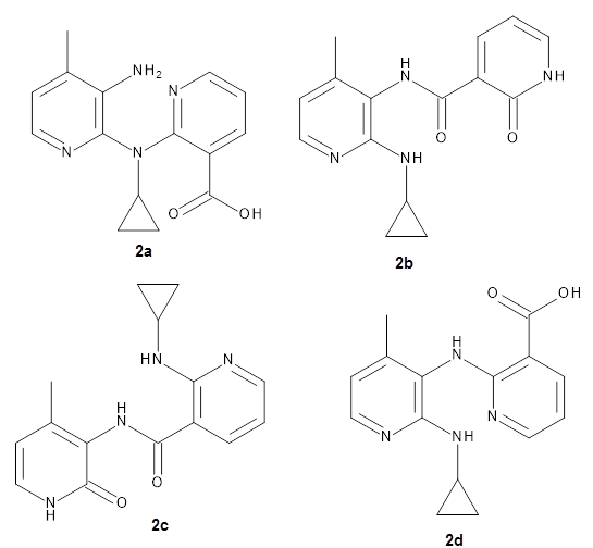
They apply the procedure to a number of test cases. For example, the HIV-1 reverse transcriptase inhibitor nevirapine hydrolyzes to a compound whose structure has been difficult to identify. The four proposed structures 2a-d were subjected to the computational method, and the 13C chemical shift RMSD for 2d is only 2.3ppm, significantly smaller than for the other 3 structures. Compound 2d was then synthesized and its NMR matches that of the nevirapine hydrolysis product.

References

1) Xin, D.; Sader, C. A.; Chaudhary, O.; Jones, P.-J.; Wagner, K.; Tautermann, C. S.; Yang, Z.; Busacca, C. A.; Saraceno, R. A.; Fandrick, K. R.; Gonnella, N. C.; Horspool, K.; Hansen, G.; Senanayake, C. H., "Development of a 13C NMR Chemical Shift Prediction Procedure Using B3LYP/cc-pVDZ and Empirically Derived Systematic Error Correction Terms: A Computational Small Molecule Structure Elucidation Method." J. Org. Chem. 2017, ASAP, DOI: 10.1021/acs.joc.7b00321.

2) Wiitala, K. W.; Hoye, T. R.; Cramer, C. J., “Hybrid Density Functional Methods Empirically Optimized for the Computation of 13C and 1H Chemical Shifts in Chloroform Solution,” J. Chem. Theory Comput. 2006, 2, 1085-1092, DOI: 10.1021/ct6001016.

3) Jensen, F., “Basis Set Convergence of Nuclear Magnetic Shielding Constants Calculated by Density Functional Methods,” J. Chem. Theory Comput., 2008, 4, 719-727, DOI: 10.1021/ct800013z.

InChIs

1: InChI=1S/C24H26F4N2O4S/c1-4-35(33,34)18-7-8-20-15(10-18)9-17(30-20)13-23(32,24(26,27)28)22(2,3)12-14-5-6-16(25)11-19(14)21(29)31/h5-11,30,32H,4,12-13H2,1-3H3,(H2,29,31)/t23-/m0/s1
InChIKey=ILKZCEOVIFOUBJ-QHCPKHFHSA-N

2d: InChI=1S/C15H16N4O2/c1-9-6-8-17-14(18-10-4-5-10)12(9)19-13-11(15(20)21)3-2-7-16-13/h2-3,6-8,10H,4-5H2,1H3,(H,16,19)(H,17,18)(H,20,21)
InChIKey=ZLFOGBWAZNUXAD-UHFFFAOYSA-N

NMR Steven Bachrach 10 Jul 2017 No Comments

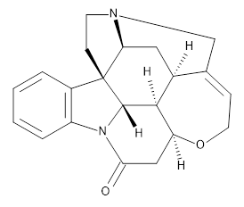
NMR coupling constants of strychnine

Helgaker, Jaszunski, and Swider1 have examined the use of B3LYP with four different basis sets to compute the spin-spin coupling constants in strychnine 1.


1

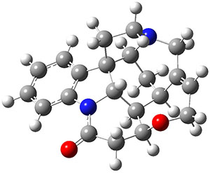
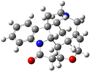
They used previously optimized coordinates of the two major conformations of strychnine, shown in Figure 1.

Conformer A

Conformer B

Figure 1. Confrmations of strychnine 1.

They tested four basis sets designed for NMR computations: pcJ-0,2 pcJ-1,2 6-31G-J,3 and 6-311G-J.3 pCJ-0 and 6-31G-J are relatively small basis sets, while the other two are considerably larger.

All four basis sets provide values of the 122 J(C-H) with a root mean square deviation of less than 0.6 Hz. J(HH) and J(CC) coupling constants are also well predicted, especially with the larger pcJ-1 basis set. They also examined the four Ramsey terms in the coupling model. The Fermi contact term dominates, and if the large pcJ-1 basis set is used to calculate it, and the smaller pcJ-0 basis set is used for the other three terms, the RMS error only increases from 0.18 to 0.20 Hz. Taking this to the extreme, they omitted calculating any of the non-Fermi contact terms, with again only small increases in the RMS – even with the small pcJ-0 basis set. Considering the computational costs, one should seriously consider whether the non-Fermi contact terms and a small basis set might be satisfactory for your own problem(s) at hand.

References

1) Helgaker, T.; Jaszuński, M.; Świder, P., "Calculation of NMR Spin–Spin Coupling Constants in Strychnine." J. Org. Chem. 2016, ASAP, DOI: 10.1021/acs.joc.6b02157.

2) Jensen, F., "The Basis Set Convergence of Spin−Spin Coupling Constants Calculated by Density Functional Methods." J. Chem. Theor. Comput. 2006, 2, 1360-1369, DOI: 10.1021/ct600166u.

3) Kjær, H.; Sauer, S. P. A., "Pople Style Basis Sets for the Calculation of NMR Spin–Spin Coupling Constants: the 6-31G-J and 6-311G-J Basis Sets." J. Chem. Theor. Comput. 2011, 7, 4070-4076, DOI: 10.1021/ct200546q.

InChIs

Strychnine 1: InChI=1S/C21H22N2O2/c24-18-10-16-19-13-9-17-21(6-7-22(17)11-12(13)5-8-25-16)14-3-1-2-4-15(14)23(18)20(19)21/h1-5,13,16-17,19-20H,6-11H2/t13-,16-,17-,19-,20-,21+/m0/s1
InChIKey=QMGVPVSNSZLJIA-FVWCLLPLSA-N

NMR Steven Bachrach 15 Nov 2016 No Comments

More examples of structure determination with computed NMR chemical shifts

Use of computed NMR chemical shifts in structure determination is really growing fast. Presented here are a couple of recent examples.

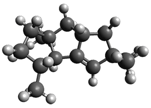
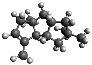
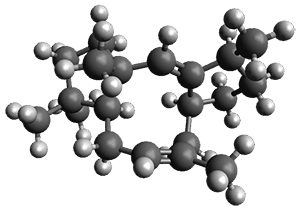
Nguyen and Tantillo used computed chemical shifts with the DP4 analysis to identify the structure of three terpenes 1-3.1 They optimized the geometries of all of the diastereomers of each compound, along with multiple conformations of each diastereomer, at B3LYP/6-31+G(d,p) and then computed the chemical shifts at SMD(CHCl3)–mPW1PW91/6-311+G(2d,p). The chemical shifts were Boltzmann weighted including all conformations within 3 kcal mol-1 of the lowest energy structure.

For 1, the DP4 analysis using just the proton shifts predicted a different isomer than using the carbon shifts, but when combined, DP4 predicted the structure, with 98.8% confidence, shown in the scheme above, and in Figure 1. For 2, the combined proton and carbon shift analysis with DP4 indicated a 100% confidence of the structure shown in the scheme and Figure 1. Lastly, for 3, which is more complicated due to the conformations of the 9-member ring, DP4 predicts with 100% confidence the structure shown in the scheme and Figure 1.

1

2

3

Figure 1. Optimized geometries of 1-3.

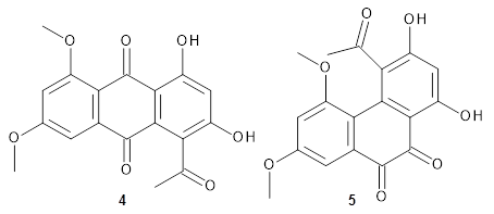
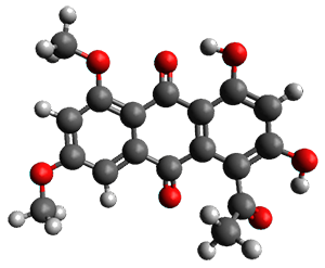
Feng, Davis and coworkers have examined a series of anthroquionones from Australian marine sponges.2 The structure of one compound was a choice of two options: 4 or 5. Initial geometries were obtain by molecular mechanics and the low energy isomers were then reoptimized at B3LYP/6-31+G(d,p). The chemical shifts were computed using PCM/MPW1PW91/6-311+G(2d,p). Application of the DP4 method indicate the structure to be 4 with a 100% confidence level. The lowest energy conformer of 4 is shown in Figure 2.

Figure 2. Optimized geometry of 4.

References

1) Nguyen, Q. N. N.; Tantillo, D. J. “Using quantum chemical computations of NMR chemical shifts to assign relative configurations of terpenes from an engineered Streptomyces host,” J. Antibiotics 2016, 69, 534–540, DOI: 10.1038/ja.2016.51.

2) Khokhar, S.; Pierens, G. K.; Hooper, J. N. A.; Ekins, M. G.; Feng, Y.; Rohan A. Davis, R. A. “Rhodocomatulin-Type Anthraquinones from the Australian Marine Invertebrates Clathria hirsuta and Comatula rotalaria,” J. Nat. Prod., 2016, 79, 946–953, DOI: 10.1021/acs.jnatprod.5b01029.

InChIs

1: InChI=1S/C15H24/c1-10-5-6-15(4)8-11-7-14(2,3)9-12(11)13(10)15/h9-11,13H,5-8H2,1-4H3/t10-,11+,13-,15+/m1/s1
InChIKey=KVSCZIPUFBVHBM-OICBVUGWSA-N

2: InChI=1S/C15H24/c1-10-5-6-15(4)8-11-7-14(2,3)9-12(11)13(10)15/h5,11-13H,6-9H2,1-4H3/t11-,12-,13+,15-/m0/s1
InChIKey=ZLYGJLHCPYVGDA-XPCVCDNBSA-N

3: InChI=1S/C20H32/c1-14-6-9-18-19(3,4)10-11-20(18,5)13-17-15(2)7-8-16(17)12-14/h6,13,15-16,18H,7-12H2,1-5H3/b14-6-,17-13-/t15-,16-,18-,20+/m0/s1
InChIKey=JZGOFJIAHJJJDK-ICZJPRMTSA-N

4: InChI=1S/C18H14O7/c1-7(19)13-10(20)6-11(21)15-16(13)17(22)9-4-8(24-2)5-12(25-3)14(9)18(15)23/h4-6,20-21H,1-3H3
InChIKey=MPQMZEXRJVMYBT-UHFFFAOYSA-N

5: InChI=1S/C18H14O7/c1-7(19)13-10(20)6-11(21)15-16(13)14-9(17(22)18(15)23)4-8(24-2)5-12(14)25-3/h4-6,20-21H,1-3H3
InChIKey=WIKIUXNPFURKNF-UHFFFAOYSA-N

NMR &terpenes Steven Bachrach 25 Oct 2016 No Comments

Further development of DP4 for NMR structure determination

Computational chemistry has had a remarkable impact on the field of structure determination by NMR spectroscopy. The ability to efficiently compute 13C and 1H chemical shifts allows for comparison of the computed chemical shifts of potential structures against the experimental values, a tremendous aid in structure determination (see some examples in previous posts). Goodman and Smith developed the DP4 method1 (see this post) to assist in identifying proper structures by means of statistical distribution of errors and Bayes Theorem.

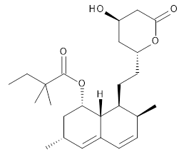
The Goodman group now reports on workflow solutions to structure prediction using DP4.2 They explore the use of open source computational tools both for predicting conformations and for computing the chemical shifts. They use a set of 10 drugs to test the performance. In general, the original DP4 method works very well in predicting drug structure, despite the fact that DP4 parameters were developed for natural products. The only failure is for simvastatin, where the large number of diastereomers and conformational flexibility prove to be too complex. The open source tools perform just slightly less effectively than the commercial packages, but are certainly a viable route for those with limited resources. The authors also provide a series of python scripts that allow users to create a seamless workflow; these should prove most helpful to the structure determination community.


Simvastatin

References

1) Smith, S. G.; Goodman, J. M. "Assigning Stereochemistry to Single Diastereoisomers by GIAO
NMR Calculation: The DP4 Probability," J. Am. Chem. Soc. 2010, 132, 12946-12959, DOI: 10.1021/ja105035r.

2) Ermanis, K.; Parkes, K. E. B.; Agback, T.; Goodman, J. M. “Expanding DP4: application to drug compounds and automation,” Org. Biomol. Chem., 2016, 14, 3943-3949, DOI: 10.1039/c6ob00015k.

InChIs

Simvastatin: InChI=1S/C25H38O5/c1-6-25(4,5)24(28)30-21-12-15(2)11-17-8-7-16(3)20(23(17)21)10-9-19-13-18(26)14-22(27)29-19/h7-8,11,15-16,18-21,23,26H,6,9-10,12-14H2,1-5H3/t15-,16-,18+,19+,20-,21-,23-/m0/s1
InChIKey=RYMZZMVNJRMUDD-HGQWONQESA-N

NMR Steven Bachrach 11 Oct 2016 No Comments

Predicting chemical structure using DP4+

Structure determination has been greatly facilitated by the use of computed NMR spectra to compare with experimental spectra. Perhaps the best method for doing this is the DP4 procedure developed by Smith and Goodman.1 (I have a previous post on their paper.) The basic idea is that if you have an experimental NMR spectrum and a number of potential structures, the computed spectra for each possibility are ranked by a statistical treatment based on the Student t-test.

Grimblat, Zanardi, and Sarotti question a couple of the assumptions embedded within the DP4 method, and offer a revision that they call DP4+.2 The two assumptions are (1) that the chemical shifts are computed at B3LYP/6-31G**//MMFF and (2) that the chemical shifts are scaled and then utilized in the analysis.

To test these assumptions, they examine a set of 72 organic compounds comprising 1219 13C shifts and 1123 1H shifts. They optimized the structures at B3LYP/6-31G* and computed the chemical shifts of these compounds using the B3LYP and mPW1PW91 functionals with 6 basis sets (6-31G*, 6-31G**, 6-31+G**, 6-311G*, 6-311G**, and 6-311+G**). With all of the combinations, the standard deviation of both the proton and carbon chemical shifts were significantly smaller than with the originally proposed method.

With regards to the second assumption, they define a new probability functions that multiplies the error using scaled chemical shifts with the error using unscaled chemical shifts, and this they call DP4+. Again with all of the computational methods, the DP4+ prediction outperforms the DP4 prediction.

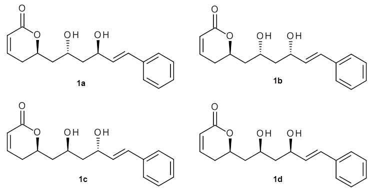
As a test case, they looked at cryptomoscatone D1 and D2 (1), for which the structures were determined with traditional methods. DP4 predicts that both cryptomoscatone D1 and D2 are structure 1d. However, DP4+ correctly predicts that cryptomoscatone D1 is 1b and cryptomoscatone D2 is 1a.

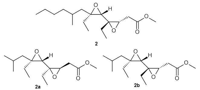
Lin and Tagliatatela-Scafati have reported the use of DP4+ to aid in the structure determination of plakdiepoxide 2.3 ROESY NMR could not provide definitive judgement of the stereochemical relationship about the bond between the two epoxide rings. They computed a number of conformers of the model compounds 2a and 2b at B3LYP/6-31G(d). The computed chemical shifts were then used with the DP4+ procedure to determine that the structure has the stereochemistry of 2b.

References

(1) Smith, S. G.; Goodman, J. M. "Assigning Stereochemistry to Single Diastereoisomers by GIAO NMR Calculation: The DP4 Probability," J. Am. Chem. Soc. 2010, 132, 12946-12959, DOI: 10.1021/ja105035r.

(2) Grimblat, N.; Zanardi, M. M.; Sarotti, A. M. "Beyond DP4: an Improved Probability
for the Stereochemical Assignment of Isomeric Compounds using Quantum Chemical Calculations of NMR Shifts," J. Org. Chem. 2015, 80, 12526-12534, DOI: 10.1021/acs.joc.5b02396.

(3) Chianese, G.; Yu, H.-B.; Yang, F.; Sirignano, C.; Luciano, P.; Han, B.-N.; Khan, S.; Lin, H.-W.; Taglialatela-Scafati, O. "PPAR Modulating Polyketides from a Chinese Plakortis simplex and Clues on the Origin of Their Chemodiversity," J. Org. Chem. 2016, 81 (12), 5135–5143, DOI: 10.1021/acs.joc.6b00695.

InChIs

Cryptomoscatone D1: InChI=1S/C17H20O4/c18-14(10-9-13-5-2-1-3-6-13)11-15(19)12-16-7-4-8-17(20)21-16/h1-6,8-10,14-16,18-19H,7,11-12H2/b10-9+/t14-,15-,16-/m1/s1
InChIKey=GOQOIZFMLWZVMB-OUUZNBFFSA-N

Cryptomoscatone D2: InChI=1S/C17H20O4/c18-14(10-9-13-5-2-1-3-6-13)11-15(19)12-16-7-4-8-17(20)21-16/h1-6,8-10,14-16,18-19H,7,11-12H2/b10-9+/t14-,15+,16+/m0/s1
InChIKey=GOQOIZFMLWZVMB-GLBZDCTLSA-N

plakdiepoxide: InChI=1S/C18H32O4/c1-6-9-10-13(4)12-17(7-2)16(22-17)18(8-3)14(21-18)11-15(19)20-5/h13-14,16H,6-12H2,1-5H3/t13?,14-,16-,17+,18-/m0/s1
InChIKey=YZQCELNTSCPAPG-KSZMJELXSA-N

NMR Steven Bachrach 20 Jun 2016 2 Comments

Dynamic effects in computing NMR (and a patent issue?)

The prediction of NMR chemical shifts and coupling constants through ab initio computation is a major development of the past decade in computational organic chemistry. I have written about many developments on this blog. An oft-used method is a linear scaling of the computed chemical shifts to match those of some test set. Kwan and Liu wondered if the dynamics of molecular motions might be why we need this correction.1

They suggest that the chemical shift can be computed as

<σ> = σ(static molecule using high level computation) + error

where the error is the obtained by using a low level computation taking the difference between the chemical shifts obtained on a dynamic molecule less that obtained with a static molecule. The dynamic system is obtained by performing molecular dynamics of the molecule, following 25 trajectories and sampling every eighth point.

They find outstanding agreement for the proton chemical shift of 12 simple molecules (mean error of 0.02 ppm) and the carbon chemical shift of 19 simple molecules (mean error of 0.5 ppm) without any scaling. Similar excellent agreement is found for a test set of natural products.

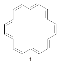
They finish up with a discussion of [18]annulene 1. The structure of 1 is controversial. X-ray crystallography indicates a near D6h geometry, but the computed NMR shifts using a D6h geometry are in dramatic disagreement with the experimental values, leading Schleyer to suggest a C2  geometry. Kwan and Liu applied their dynamic NMR method to the D6h, D3h, and C2 structures, and find the best agreement with the experimental chemical shifts are from the dynamic NMR initiated from the D6h geometry. Dynamic effects thus make up for the gross error found with the static geometry, and now bring the experimental and computational data into accord.

One final note on this paper. The authors indicate that they have filed a provisional patent on their method. I am disturbed by this concept of patenting a computational methodology, especially in light of the fact that many other methods have been made available to the world without any legal restriction. For example, full details including scripts to apply Tantillo’s correction method are available through the Cheshire site and a web app to implement Goodman’s DP4 method are available for free. Provisional patents are not available for review from the US Patent Office so I cannot assess just what is being protected here. However, I believe that this action poses a real concern over the free and ready exchange of computational methodologies and ideas.

References

(1) Kwan, E. E.; Liu, R. Y. "Enhancing NMR Prediction for Organic Compounds Using Molecular Dynamics," J. Chem. Theor. Comput. 2015, 11, 5083-5089, DOI: 10.1021/acs.jctc.5b00856.

InChIs

1: InChI=1S/C18H18/c1-2-4-6-8-10-12-14-16-18-17-15-13-11-9-7-5-3-1/h1-18H/b2-1-,3-1+,4-2+,5-3+,6-4+,7-5-,8-6-,9-7+,10-8+,11-9+,12-10+,13-11-,14-12-,15-13+,16-14+,17-15+,18-16+,18-17-
InChIKey=STQWAGYDANTDNA-DWSNDWDZSA-N

NMR Steven Bachrach 11 Jan 2016 4 Comments

Structure revision: Vescalagin and Castalagin

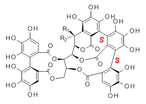
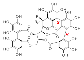
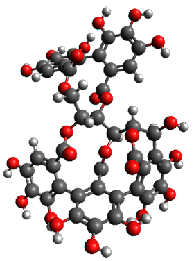
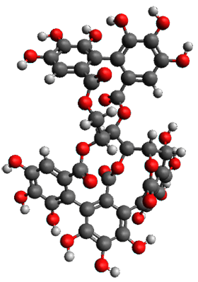
Vescalagin 1 and castalagin 2 are found in plants and also in wine and whisky. They possess some intriguing stereochemistry and the topic of interest in the paper by Tanaka and coworkers is the stereochemistry of the triphenyl fragment.1 The original proposed structure indicated a (S,S) (1a and 2a) configuration, yet a molecular mechanics study suggest the (S,R) (1b and 2b) configuration would be lower in energy.

1a: R1 = OH, R2 = H
2a: R1 = H, R2 = OH

1b: R1 = OH, R2 = H
2b: R1 = H, R2 = OH

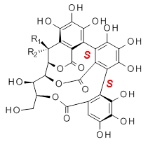
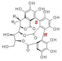
Recognizing the power of DFT computations in resolving this type of structural problem, Tanaka measured the ECD spectrum of the hydrolyzed forms of 1 and 2, namely 3 and 4. The (S,S) and (S,R) isomers of 3 and 4 were subjected to a Monte Carlo search using MM. Low-lying conformers were reoptimized at B3LYP/6-31G(d,p) including PCM, modeling methanol as the solvent. The ECD spectrum was then predicted using all conformations with a population over 1%. The computed spectrum for the (S,R) isomer reproduced the negative Cotton effect at 218 nm observed in the experiment.

3a: R1 = OH, R2 = H
4a: R1 = H, R2 = OH

3b: R1 = OH, R2 = H
3b: R1 = H, R2 = OH

The structures of 1 and 2 of both stereoisomers were next optimized at B3LYP/6-31G(d,p) including PCM. The lowest energy conformation of each is shown in Figure 1. The 1H and 13C chemical shifts were computed at this level, again using all conformations with a population greater than 1%. The correlation coefficient for the fit between the experimental values of the chemical shifts and 1a and 2a are significantly lower for both proton and carbon, while the correlation coefficients compared to 1b and 2b are larger, 0.93 or better. Therefore, the structures of vescalagin is 1b and castalagin is 2b.

1b

2b

Figure 1. B3LYP/6-31G(d,p) optimized geometries of the lowest energy conformers of 1b and 2b.

References

(1) Matsuo, Y.; Wakamatsu, H.; Omar, M.; Tanaka, T. "Reinvestigation of the Stereochemistry of the C-Glycosidic Ellagitannins, Vescalagin and Castalagin," Org. Lett. 2014, 17, 46-49, DOI: 10.1021/ol503212v.

InChIs

1: InChI=1S/C41H26O26/c42-8-1-5-12(24(48)21(8)45)13-6(2-9(43)22(46)25(13)49)39(60)65-34-11(4-63-37(5)58)64-38(59)7-3-10(44)23(47)26(50)14(7)15-18-16(28(52)32(56)27(15)51)17-19-20(30(54)33(57)29(17)53)31(55)35(66-41(19)62)36(34)67-40(18)61/h1-3,11,31,34-36,42-57H,4H2/t11-,31-,34+,35+,36-/m0/s1
InChIKey=UDYKDZHZAKSYCO-KWVBPWBCSA-N

2: InChI=1S/C41H26O26/c42-8-1-5-12(24(48)21(8)45)13-6(2-9(43)22(46)25(13)49)39(60)65-34-11(4-63-37(5)58)64-38(59)7-3-10(44)23(47)26(50)14(7)15-18-16(28(52)32(56)27(15)51)17-19-20(30(54)33(57)29(17)53)31(55)35(66-41(19)62)36(34)67-40(18)61/h1-3,11,31,34-36,42-57H,4H2/t11-,31+,34+,35+,36-/m0/s1
InChIKey=UDYKDZHZAKSYCO-GJTMBUPBSA-N

NMR Steven Bachrach 09 Mar 2015 No Comments

Next Page »