Archive for the 'nucleic acids' Category

Gas phase structure of uridine

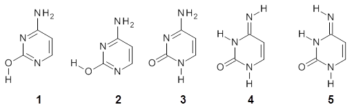
To advance our understanding of why ribose takes on the furanose form, rather than the pyranose form, in RNA, Alonso and co-workers have examined the structure of uridine 1 in the gas phase.1
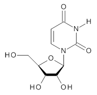

1

Uridine is sensitive to temperature, and so the laser-ablation method long used by the Alonso group is ideal for examining uridine. The microwave spectrum is quite complicated due to the presence of many photofragments. Careful analysis lead to the identification of a number of lines and hyperfine structure that could be definitively assigned to uridine, leading to experimental values of the rotational constants and the diagonal elements of the 14N nuclear quadrupole coupling tensor for each nitrogen. These values are listed in Table 1.

Table 1. Experimental and calculated rotational constants (MHz), quadrupole coupling constants (MHz) and relative energy (kcal mol-1).


calculated


Expt.

anti/C2’-endo-g+

syn/C2’-endo-g+

anti/C3’-endo-g+

anti/C2’-endo-t

syn/C3’-endo-g+

A

885.98961

901.2

935.8

790.0

799.7

925.5

B

335.59622

340.6

308.4

352.6

330.6

300.4

C

270.11210

276.6

266.6

261.4

262.9

264.0

14N1 χxx

1.540

1.50

1.82

1.48

1.46

1.82

14N1 χyy

1.456

1.43

0.73

1.71

1.81

-0.72

14N1 χzz

-2.996

-2.93

-2.56

-3.19

-3.27

-1.11

14N3 χxx

1.719

1.74

2.03

1.78

1.62

1.98

14N3 χyy

1.261

1.11

0.47

1.34

1.51

-0.75

14N3 χzz

-2.979

-2.85

-2.50

-3.12

-3.13

-1.23

Rel E


0.0

1.10

1.90

2.00

2.15

In order to assign a 3-D structure to these experimental values, they examined the PES of uridine with molecular mechanics and semi-empirical methods, before reoptimizing the structure of the lowest 5 energy structures at MP2/6-311++G(d,p). Then, comparison of the resulting rotational constants and 14N nuclear quadrupole coupling constants of these computed structures (see Table 1) led to identification of the lowest energy structure (anti/C2’-endo-g+, see Figure 1) in best agreement with the experiment. Once again, the Alonso group has demonstrated the value of the synergy between experiment and computation in structure identification.

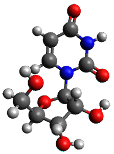
Figure 1. MP2/6-311++G(d,p) optimized structure of 1 (anti/C2’-endo-g+).

References

(1) Peña, I.; Cabezas, C.; Alonso, J. L. "The Nucleoside Uridine Isolated in the Gas Phase," Angew. Chem. Int. Ed. 2015, 54, 2991-2994, DOI: 10.1002/anie.201412460.

Inchis:

1: Inchi=1S/C9H12N2O6/c12-3-4-6(14)7(15)8(17-4)11-2-1-5(13)10-9(11)16/h1-2,4,6-8,12,14-15H,3H2,(H,10,13,16)/t4-,6-,7-,8-/m1/s1
InChiKey=DRTQHJPVMGBUCF-XVFCMESISA-N

MP &nucleic acids Steven Bachrach 06 Apr 2015 No Comments

Gas-phase structure of cytosine

Alonso and coworkers have again (see this post employed laser-ablation molecular-beam Fourier-transform microwave (LA-MB-MW)spectroscopy to discern the gas phase structure of an important biological compound: cytosine.1 They identified five tautomers of cytosine 1-5. Comparison between the experimental and computational (MP2/6-311++G(d,p) microwave rotational constants and nitrogen nuclear quadrupole coupling constants led to the complete assignment of the spectra. The experimental and calculated rotational constants are listed in Table 1.

Table 1. Rotational constants (MHz) for 1-5.


1

2

3

4

5


Expt

calc

Expt

calc

Expt

calc

Expt

calc

Expt

calc

A

3951.85

3934.5

3889.46

3876.5

3871.55

3856.0

3848.18

3820.1

3861.30

3844.2

B

2008.96

1999.1

2026.32

2014.7

2024.98

2012.3

2026.31

2019.0

2011.41

1999.7

C

1332.47

1326.8

1332.87

1326.9

1330.34

1323.3

1327.99

1324.0

1323.20

1318.4

The experimental and computed relative free energies are listed in Table 2. There is both not a complete match of the relative energetic ordering of the tautomers, nor is there good agreement in their magnitude. Previous computations2 at CCSD(T)/cc-pVQZ//CCSD//cc-pVTZ are in somewhat better agreement with the gas-phase experiments.

Table 2. Relative free energies (kcal mol-1) of 1-5.


expt

MP2/
6-311++G(d,p)

CCSD(T)/cc-pVQZ//
CCSD//cc-pVTZ

1

0.0

0.0

0.0

2

0.47

0.70

0.7

3

0.11

1.19

0.2

4

0.83

3.61

0.7

5


5.22


References

(1) Alonso, J. L.; Vaquero, V.; Peña, I.; López, J. C.; Mata, S.; Caminati, W. "All Five Forms of Cytosine Revealed in the Gas Phase," Angew. Chem. Int. Ed. 2013, 52, 2331-2334, DOI: 10.1002/anie.201207744.

(2) Bazso, G.; Tarczay, G.; Fogarasi, G.; Szalay, P. G. "Tautomers of cytosine and their excited electronic states: a matrix isolation spectroscopic and quantum chemical study," Phys. Chem. Chem. Phys., 2011, 13, 6799-6807, DOI:10.1039/C0CP02354J.

InChIs

cytosine: InChI=1S/C4H5N3O/c5-3-1-2-6-4(8)7-3/h1-2H,(H3,5,6,7,8)
InChIKey=OPTASPLRGRRNAP-UHFFFAOYSA-N

MP &nucleic acids Steven Bachrach 22 Apr 2013 1 Comment

Gaunine tautomers

Here’s another fine paper from the Alonso group employing laser ablation molecular beam Fourier transform microwave spectroscopy coupled with computation to discern molecular structure. In this work they examine the low-energy tautomers of guanine.1 The four lowest energy guanine tautomers are shown in Figure 1. (Unfortunately, Alonso does not include the optimized coordinates of these structures in the supporting information – we need to more vigorously police this during the review process!) These tautomers are predicted to be very close in energy (MP2/6-311++G(d,p), and so one might expect to see multiple signals in the microwave originating from all four tautomers. In fact, they discern all four, and the agreement between the computed and experimental rotational constants are excellent (Table 1), especially if one applies a scaling factor of 1.004. Once again, this group shows the power of combined experiment and computations!


1 (0.0)


2 (0.28)


3 (0.40)


4 (0.99)

Figure 1. Four lowest energy (kcal mol-1, MP2/6-311++G(d,p)) tautomers of guanine.

Table 1. Experimental and computed rotational constants (MHz) of the four guanine tautomers.


1

2

3

4


Exp

Comp

Exp

Comp

Exp

Comp

Exp

Comp

A

19.22155

1909.0

19.222780

1909.7

1916.080

1908.6

1923.460

1915.6

B

1121.6840

119.2

1116.6710

1113.5

1132.360

1128.2

1136.040

1131.9

C

709.0079

706.6

706.8580

704.2

712.1950

709.5

714.7000

712.0

References

(1) Alonso, J. L.; Peña, I.; López, J. C.; Vaquero, V., "Rotational Spectral Signatures of Four Tautomers of Guanine," Angew. Chem. Int. Ed. 2009, 48, 6141-6143, DOI: 10.1002/anie.200901462

InChIs

Guanine: InChI=1/C5H5N5O/c6-5-9-3-2(4(11)10-5)7-1-8-3/h1H,(H4,6,7,8,9,10,11)/f/h8,10H,6H2
InChIKey=UYTPUPDQBNUYGX-GSQBSFCVCX

MP &nucleic acids Steven Bachrach 05 Oct 2009 3 Comments

DFT performance with nucleic acid base pairs

Here is another benchmark of the performance of DFT in handling difficult situations, in this case the interaction between nucleic acid base pairs. Sherrill1 has examined the 124 nucleic acid base pairs from the JSCH-2005 database2 compiled by Hobza and coworkers. This database includes 36 hydrogen bonded complexes, and example of which is shown in Figure 1a, and 54 stacked complex, one example of which is shown in Figure 1b.

(a)

(b)

Figure 1. Optimized geometries (RI-MP2/cc-pVTZ) of two representative structures of base pairs: (a) hydrogen bonded pair and (c) stacked pair.

The energies of these base pairs computed with four different functionals: PBE, PBE-D (where Grimme’s empirical dispersion correction3), and the recently developed MO5-2X4 and MO6-2X5 methods which attempt to treat mid-range electron correlation. The aug-cc-pVDZ basis set was used. These DFT energies are compared with the CCSD(T) energies of Hobza. The mean unsigned error (MUE) for the 28 hydrogen bonded complexes and the 54 stacked complexes are listed in Table 1.

Table 1. Mean unsigned error (kcal mol-1) of the four DFT
methods (relative to CCSD(T)) for the hydrogen bonded and stacked base pairs.


method

MUE (HB)

MUE (stacked)


PBE

2.59

7.57

PBE-D

0.70

1.53

MO5-2X

1.98

2.59

MO6-2X

1.62

1.08


A few interesting trends are readily apparent. First, PBE (representing standard GGA DFT methods) poorly describes the energy of the hydrogen bonded complexes, but utterly fails to treat the stacking interaction. Inclusion of the dispersion correction (PBE-D) results in excellent energies for the HB cases and quite reasonable results for the stacked pairs. Both of Truhlar’s functionals dramatically outperform PBE, though MO5-2X is probably still not appropriate for the stacked case. MO6-2X however seems to be a very reasonable functional for dealing with base pair interactions, indicating that mid-range correlation correction is sufficient to describe these complexes, and that the long-range correlation correction included in the dispersion correction, while giving improved results, is not essential.

References

(1) Hohenstein, E. G.; Chill, S. T.; Sherrill, C. D., "Assessment of the Performance of the M05-2X and M06-2X Exchange-Correlation Functionals for Noncovalent Interactions in Biomolecules," J. Chem. Theory Comput., 2008, 4, 1996-2000, DOI: 10.1021/ct800308k

(2) Jurecka, P.; Sponer, J.; Cerny, J.; P., H., "Benchmark database of accurate (MP2 and CCSD(T) complete basis set limit) interaction energies of small model complexes, DNA base pairs, and amino acid pairs," Phys. Chem. Chem. Phys., 2006, 8, 1985-1993, DOI: 10.1039/b600027d.

(3) Grimme, S., "Semiempirical GGA-type density functional constructed with a long-range dispersion correction," J. Comput. Chem., 2006, 27, 1787-1799, DOI: 10.1002/jcc.20495

(4) Zhao, Y.; Schultz, N. E.; Truhlar, D. G., "Design of Density Functionals by Combining the Method of Constraint Satisfaction with Parametrization for Thermochemistry, Thermochemical Kinetics, and Noncovalent Interactions," J. Chem. Theory Comput., 2006, 2, 364-382, DOI: 10.1021/ct0502763.

(5) Zhao, Y.; Truhlar, D. G., "The M06 suite of density functionals for main group thermochemistry, thermochemical kinetics, noncovalent interactions, excited states, and transition elements: two new functionals and systematic testing of four M06-class functionals and 12 other functionals," Theor. Chem. Acc., 2008, 120, 215-241, DOI: 10.1007/s00214-007-0310-x.

DFT &nucleic acids Steven Bachrach 12 Jan 2009 2 Comments