Archive for the 'Enzyme' Category

Three interesting recent Angew. Chem. papers

A note here on a few recent Angew. Chem. articles of interest to readers of this blog. The first is a comment by Frenking1 concerning the “trilogue” by Shaik, Hoffmann and Rzepa2 which discusses the nature of C2, especially the notion that this molecules may possess a quadruple bond (see this post for a previous post on this article.) Frenking argues that the force constant associated with the C-C stretch in C2 is smaller than that in acetylene, so how can one argue that there is some quadruple bond character in C2? A reply from the original authors3 accompanies the comment by Frenking, and they respond by noting that the PES for bond stretching is unusually flat. I had the generally sense, though, that the authors of both articles were really talking past each other and that an opportunity for a more fruitful discussion has been missed.

The other article of note is an excellent review of de novo enzyme design as performed by the Baker and Houk labs.4 This review, authored by leaders of this effort, highlights their approach to this “holy grail” problem. The general notion is to use standard tools of computational chemistry to design a theozyme. Next, this theozyme is placed into known protein motifs with the attempt to have it fit without too much steric clash. The protein is then mutated one residue at a time to optimize the fit and binding of the theozyme to substrate. Lastly, the best targets are synthesized and tested. (The reader can see my post one of their projects: synthetic Diels-Alderase.)

References

(1) Frenking, G.; Hermann, M. "Critical Comments on “One Molecule, Two Atoms, Three
Views, Four Bonds?”," Angew. Chem. Int. Ed. 2013, 52, 5922-5925. DOI: 10.1002/anie.201301485.

(2) Shaik, S.; Rzepa, H. S.; Hoffmann, R. "One Molecule, Two Atoms, Three Views, Four
Bonds?," Angew. Chem. Int. Ed. 2013, 52, 3020-3033, DOI: 10.1002/anie.201208206.

(3) Danovich, D.; Shaik, S.; Rzepa, H. S.; Hoffmann, R. "A Response to the Critical Comments on “One Molecule, Two Atoms, Three Views, Four Bonds?”," Angew. Chem. Int. Ed. 2013, 52, 5926-5928, DOI: 10.1002/anie.201302350.

(4) Kiss, G.; Çelebi-Ölçüm, N.; Moretti, R.; Baker, D.; Houk, K. N. "Computational Enzyme Design," Angew. Chem. Int. Ed. 2013, 52, 5700-5725, DOI: 10.1002/anie.201204077.

Bond Dissociation Energy &Enzyme Steven Bachrach 24 Jun 2013 No Comments

Enzymatic catalysis of ladder ether formation

Biosynthesis of ladder polyethers is the topic of a very nice experimental/computational study by Chen and Houk.1 The x-ray structure of the enzyme that catalyzes the nucleophilic attack on epoxides to create the 6-member ring ether was determined, but the geometry did not completely indicate the mechanism.

Gas phase computations of the 5-exo-tet and 6-endo-tet ring openings of 1 were examined for both the acid and base catalyzed routes at B2PLYP/6-311++G(d,p)//B2PLYP/6-31G(d).
The results are summarized in Figure 1. Basically, as expected by Baldwin’s rules, the closure to the tetrahydrofuran (5-exo-tet) is favored under both catalyzed conditions. However, the preference is small under base conditions, with the difference in the free energy of activation of only 1.2 kcal mol-1.

Figure 1. Gas phase energies (kcal mol-1) for the acid an base catalyzed reactions of 1 to 2 or 3.

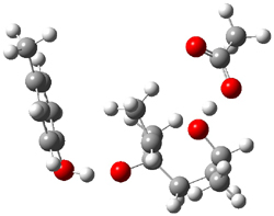
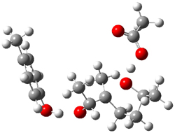
The enzyme Lsd19B produces just the analogue of 3. So, the two regioisomeric TSs were reoptimized with an aspartic acid group and a tyrosine group in the positions they occupy in the active site of the enzyme Lsd19b. The two resulting transition states, evaluated at B2LYP/6-311++G(d,p)//MO6-2x/6-31G(d), are shown in Figure 2. The activation energy for the 6-endo-tet reaction is 18.0 kcal mol-1, 2.5 kcal mol-1 lower than for the 5-exo-tet route. This energy difference would give rise to a 100:1 selectivity for the tetrahydropyran product, in accord with experiment. The enzyme preorganizes for and favors the base catalyzed path that leads to 3.

5-exo-tet TS model
ΔG‡ = 20.5

6-endo-tet TS model
ΔG‡ = 18.0

Figure 2. Transition state models of the active site. Activation energies in kcal mol-1.

References

(1) Hotta, K.; Chen, X.; Paton, R. S.; Minami, A.; Li, H.; Swaminathan, K.; Mathews, I. I.; Watanabe, K.; Oikawa, H.; Houk, K. N.; Kim, C.-Y., "Enzymatic catalysis of anti-Baldwin ring closure in polyether biosynthesis," Nature, 2012, 483, 355-358, DOI: 10.1038/nature10865.

InChIs

1: InChI=1/C8H16O2/c1-6(9)4-5-8(3)7(2)10-8/h6-7,9H,4-5H2,1-3H3/t6-,7?,8+/m0/s1
InChIKey=YEAGKBPXLVGCPK-YPVSKDHRBB

2: InChI=1/C8H16O2/c1-6-4-5-8(3,10-6)7(2)9/h6-7,9H,4-5H2,1-3H3/t6-,7-,8-/m0/s1
InChIKey=RRDMOYCGUXNJSL-FXQIFTODBG

3: InChI=1/C8H16O2/c1-6-4-5-8(3,9)7(2)10-6/h6-7,9H,4-5H2,1-3H3/t6-,7-,8+/m0/s1
InChIKey=WWLWIBPPUNCAQU-BIIVOSGPBQ

Enzyme Steven Bachrach 24 Apr 2012 1 Comment

Designing a Diels-Alderase

One of the great challenges to computational chemistry and computational biochemistry is rational design of enzymes. Baker and Houk have been pursuing this goal and in their recent paper they report progress towards an enzyme designed to catalyze a Diels-Alder reaction.1

They envisaged an enzyme that could catalyze the Diels-Alder of 1 with 2 by having a suitable hydrogen bond acceptor of the carbamide proton of 1 (such as the carbonyl oxygen of glutamine or asparagine) along with a suitable donor to the oxygen of 2 (such as the hydroxyl of tyrosine, serine or threonine) – as shown below. Along with positioning the diene and dienophile near each other and properly orienting them for reaction, the activation barrier should be lowered by narrowing the HOMO-LUMO gap.

A series of transition states for the Diels-Alder reaction of 1 with 2 along with the hydrogen-bonded amino acids were optimized B3LYP/6-31+G(d,p) and used as constraints within the RosettaMatch code for locating a protein scaffold that could accommodate this TS structure. This resulted in 84 protein designs, each of which were synthesized and screened for activity in catalyzing the Diels-Alder reaction. Of these potential enzymes, 50 were soluble and of these 50, only 2 showed any activity. These two were selectively mutated to try to improve activity, and some improvement was obtained.

Of particular note is that mutation that removed one or both of the residues designed to hydrogen bond to the substrates resulted in complete loss of activity.

In principle 8 different steriosomeric products are possible in the reaction of 1 with 2. In solution in the absence of enzyme, four products are observed, with the major product (47%) the 3R,4S endo prodcut 3. The designed enzymes were constructed to make this product, and in fact it is the only observed stereoisomer formed in the reaction in the presence of enzyme. Furthermore, the designed enzymes are quite selective; for example, changing a single N-methyl group to N-ethyl on 2 reduced the rate by a factor of 2 and larger substituents resulted in a greater rate suppression.

Turnover rate is high and suggests that these enzymes might have real application in chemical synthesis. The disappointing aspect of the study was the poor ratio of predicted enzymes (84) to ones that actually had activity (2).

References

(1) Siegel, J. B.; Zanghellini, A.; Lovick, H. M.; Kiss, G.; Lambert, A. R.; St.Clair, J. L.; Gallaher, J. L.; Hilvert, D.; Gelb, M. H.; Stoddard, B. L.; Houk, K. N.; Michael, F. E.; Baker, D., "Computational Design of an Enzyme Catalyst for a Stereoselective Bimolecular Diels-Alder Reaction," Science, 2010, 329, 309-313, DOI: 10.1126/science.1190239

Enzyme &Houk Steven Bachrach 18 Jan 2012 2 Comments

New enzyme activation model

The standard model for explaining enzyme activation is that the active site is designed to stabilize the transition state, thereby reducing the activation barrier. Jonathan Goodman offers a very compelling argument for an alternative explanation for at least some enzymes.1

He examined enzymes that coordinate the substrate through what’s called an “oxyanion hole”, a region in the active site where an incipient oxyanion can be stabilized through 2 or three hydrogen bonds. This usually involves nucleophilic attack at a carbonyl. Analysis of the protein data bank turned up several hundred such structures where a carbonyl is coordinated to the enzyme by 2 or more hydrogen bonds. Also examined were several hundred small molecule x-ray structures that also exhibit this sort of hydrogen bonding scheme. The geometry about the carbonyl oxygen was examined – distances angles and dihedral angles – and the only significant difference between the enzyme and small molecule set is for the dihedral angle formed between the O=C-R plane of the carbonyl and the C=OH angle to the hydrogen bond donor. For the small molecules, the preferred value is about 0°, but for the enzymes, the preferred angle is about 90°.

MPWB1K/6-311++G**//B3LYP/6-31G(d,p) computations of a model enzyme active site (see Scheme 1) were performed where the two waters are arranged at different dihedral angles. For both reactant and transition state, the coordinating waters stabilize the structures – and there is a stabilization for all dihedral angles.

Scheme 1

But the best arrangement, i.e. the maximum stabilization, occurs when the waters are arranged with a dihedral angle of 0° for both the reactant and transition state. At 0°, the reactant is significantly stabilized, more so than the stabilization of the TS. At 90° stabilization of both species is less than at 0° but the stabilization is much less for the reactant than for the TS. Thus, at 90° the activation barrier is lowered not by preferential stabilization of the TS but by lesser stabilization of the reactant! The active site is set up not to stabilize the TS but rather to minimize the activation barrier through differential stabilization of the reactant vs the TS. This new model offer another approach towards creating artificial catalysts, ones designed not to maximize binging, but rather to minimize the activation barrier through judicious stabilization of the TS and destabilization of the reactant.

References

(1) Simon, L.; Goodman, J. M., "Enzyme Catalysis by Hydrogen Bonds: The Balance between Transition State Binding and Substrate Binding in Oxyanion Holes," J. Org. Chem. 2010, DOI: 10.1021/jo901503d

Enzyme Steven Bachrach 18 Jan 2010 1 Comment