Archive for the 'MP' Category

Long C-C bonds are not caused by crystal packing forces

Schreiner and Grimme have examined a few compounds (see these previous posts) with long C-C bonds that are found in congested systems where dispersion greatly aids in stabilizing the stretched bond. Their new paper1 continues this theme by examining 1 (again) and 2, using computations, and x-ray crystallography and gas-phase rotational spectroscopy and electron diffraction to establish the long C-C bond.

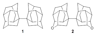
The distance of the long central bond in 1 is 1.647 Å (x-ray) and 1.630 Å (electron diffraction). Similarly, this distance in 2 is 1.642 Å (x-ray) and 1.632 Å (ED). These experiments discount any role for crystal packing forces in leading to the long bond.

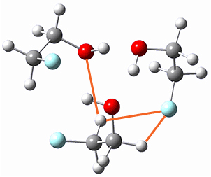
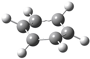
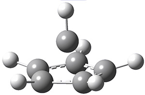
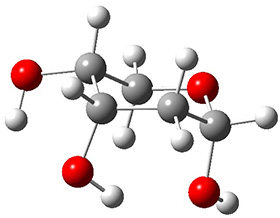
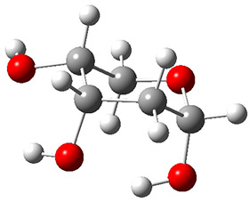
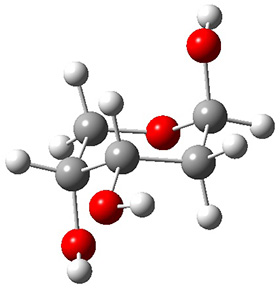
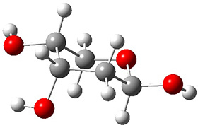
A very nice result from the computations is that most functionals that include some dispersion correction predict the C-C distance in the optimized structures with an error of no more than 0.01 Å. (PW6B95-D3/DEF2-QZVP structures are shown in Figure 1.) Not surprisingly, HF and B3LYP without a dispersion correction predict a bond that is too long.) MP2 predicts a distance that is too short, but SCS-MP2 does a very good job.


1


2

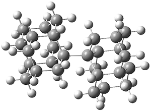
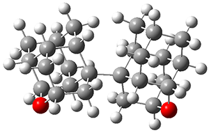
Figure 1. PW6B95-D3/DEF2-QZVP optimized structures of 1 and 2.

References

1) Fokin, A. A.; Zhuk, T. S.; Blomeyer, S.; Pérez, C.; Chernish, L. V.; Pashenko, A. E.; Antony, J.; Vishnevskiy, Y. V.; Berger, R. J. F.; Grimme, S.; Logemann, C.; Schnell, M.; Mitzel, N. W.; Schreiner, P. R., "Intramolecular London Dispersion Interaction Effects on Gas-Phase and Solid-State Structures of Diamondoid Dimers." J. Am. Chem. Soc. 2017, 139, 16696-16707, DOI: 10.1021/jacs.7b07884.

InChIs

1: InChI=1S/C28H38/c1-13-7-23-19-3-15-4-20(17(1)19)24(8-13)27(23,11-15)28-12-16-5-21-18-2-14(9-25(21)28)10-26(28)22(18)6-16/h13-26H,1-12H2
InChIKey=MMYAZLNWLGPULP-UHFFFAOYSA-N

2: InChI=1S/C26H34O2/c1-11-3-19-15-7-13-9-25(19,21(5-11)23(27-13)17(1)15)26-10-14-8-16-18-2-12(4-20(16)26)6-22(26)24(18)28-14/h11-24H,1-10H2
InChIKey=VPBJYHMTINJMAE-UHFFFAOYSA-N

adamantane &DFT &Grimme &MP &Schreiner Steven Bachrach 25 Jun 2018 1 Comment

Structure of the 2-fluoroethanol trimer

Here is another fine example of the power of combining experiment and computation. Xu and co-worker has applied the FT microwave technique, which has been used in conjunction with computation by the Alonso group (especially) as described in these posts, to the trimer of 2-fluoroethanol.1 They computed a number of trimer structures at MP2/6-311++G(2d,p) in an attempt to match up the computed spectroscopic constants with the experimental constants. The two lowest energy structures are shown in Figure 1. The second lowest energy structure has nice symmetry, but it does not match up well with the experimental spectra. However, the lowest energy structure is in very good agreement with the experiments.

(0.0)

(4.15)

Table 1. MP2/6-311++G(2d,p) optimized structures and relative energies (kJ mol-1) of the two lowest energy structures of the trimer of 2-fluoroethanol. The added orange lines in the lowest energy structure denote the bifurcated hydrogen bonds identified by QTAIM.

Of particular note is that topological electron density analysis (also known as quantum theoretical atoms in a molecule, QTAIM) of the wavefunction of the lowest energy structure of the trimer identifies two hydrogen bond bifurcations. The authors suggest that these additional interactions are responsible, in part, for the stability of this lowest energy structure.

References

(1) Thomas, J.; Liu, X.; Jäger, W.; Xu, Y. "Unusual H-Bond Topology and Bifurcated H-bonds in the 2-Fluoroethanol Trimer," Angew. Chem. Int. Ed. 2015, 54, 11711-11715, DOI: 10.1002/anie.201505934.

InChIs

2-fluoroethanol: InChI=1S/C2H5FO/c3-1-2-4/h4H,1-2H2, InChIKey=GGDYAKVUZMZKRV-UHFFFAOYSA-N

Hydrogen bond &MP Steven Bachrach 20 Oct 2015 1 Comment

Structure of 2-oxazoline

A recent reinvestigation of the structure of 2-oxazoline demonstrates the difficulties that many computational methods can still have in predicting structure.

Samdal, et al. report the careful examination of the microwave spectrum of 2-oxzoline and find that the molecule is puckered in the ground state.1 It’s not puckered by much, and the barrier for inversion of the pucker, through a planar transition state is only 49 ± 8 J mol-1. The lowest vibrational frequency in the non-planar ground state, which corresponds to the puckering vibration, has a frequency of 92 ± 15 cm-1. This low barrier is a great test case for quantum mechanical methodologies.

And the outcome here is not particularly good. HF/cc-pVQZ, M06-2X/cc-pVQZ, and B3LYP/cc-pVQZ all predict that 2-oxazoline is planar. More concerning is that CCSD and CCSD(T) with either the cc-pVTZ or cc-pVQZ basis sets also predict a planar structure. CCSD(T)-F12 with the cc-pVDZ predicts a non-planar ground state with a barrier of only 8.5 J mol-1, but this barrier shrinks to 5.5 J mol-1 with the larger cc-pVTZ basis set.

The only method that has good agreement with experiment is MP2. This method predicts a non-planar ground state with a pucker barrier of 11 J mol-1 with cc-pVTZ, 39.6 J mol-1 with cc-pVQZ, and 61 J mol-1 with the cc-pV5Z basis set. The non-planar ground state and the planar transition state of 2-oxazoline are shown in Figure 1. The computed puckering vibrational frequency does not reproduce the experiment as well; at MP2/cc-pV5Z the predicted frequency is 61 cm-1 which lies outside of the error range of the experimental value.

Non-planar

Planar TS

Figure 1. MP2/cc-pV5Z optimized geometry of the non-planar ground state and the planar transition
state of 2-oxazoline.

References

(1) Samdal, S.; Møllendal, H.; Reine, S.; Guillemin, J.-C. "Ring Planarity Problem of 2-Oxazoline Revisited Using Microwave Spectroscopy and Quantum Chemical Calculations," J. Phys. Chem. A 2015, 119, 4875–4884, DOI: 10.1021/acs.jpca.5b02528.

InChIs

2-oxazoline: InChI=1S/C3H5NO/c1-2-5-3-4-1/h3H,1-2H2
InChIKey=IMSODMZESSGVBE-UHFFFAOYSA-N

MP &vibrational frequencies Steven Bachrach 15 Jun 2015 1 Comment

Gas phase structure of uridine

To advance our understanding of why ribose takes on the furanose form, rather than the pyranose form, in RNA, Alonso and co-workers have examined the structure of uridine 1 in the gas phase.1


1

Uridine is sensitive to temperature, and so the laser-ablation method long used by the Alonso group is ideal for examining uridine. The microwave spectrum is quite complicated due to the presence of many photofragments. Careful analysis lead to the identification of a number of lines and hyperfine structure that could be definitively assigned to uridine, leading to experimental values of the rotational constants and the diagonal elements of the 14N nuclear quadrupole coupling tensor for each nitrogen. These values are listed in Table 1.

Table 1. Experimental and calculated rotational constants (MHz), quadrupole coupling constants (MHz) and relative energy (kcal mol-1).


calculated


Expt.

anti/C2’-endo-g+

syn/C2’-endo-g+

anti/C3’-endo-g+

anti/C2’-endo-t

syn/C3’-endo-g+

A

885.98961

901.2

935.8

790.0

799.7

925.5

B

335.59622

340.6

308.4

352.6

330.6

300.4

C

270.11210

276.6

266.6

261.4

262.9

264.0

14N1 χxx

1.540

1.50

1.82

1.48

1.46

1.82

14N1 χyy

1.456

1.43

0.73

1.71

1.81

-0.72

14N1 χzz

-2.996

-2.93

-2.56

-3.19

-3.27

-1.11

14N3 χxx

1.719

1.74

2.03

1.78

1.62

1.98

14N3 χyy

1.261

1.11

0.47

1.34

1.51

-0.75

14N3 χzz

-2.979

-2.85

-2.50

-3.12

-3.13

-1.23

Rel E


0.0

1.10

1.90

2.00

2.15

In order to assign a 3-D structure to these experimental values, they examined the PES of uridine with molecular mechanics and semi-empirical methods, before reoptimizing the structure of the lowest 5 energy structures at MP2/6-311++G(d,p). Then, comparison of the resulting rotational constants and 14N nuclear quadrupole coupling constants of these computed structures (see Table 1) led to identification of the lowest energy structure (anti/C2’-endo-g+, see Figure 1) in best agreement with the experiment. Once again, the Alonso group has demonstrated the value of the synergy between experiment and computation in structure identification.

Figure 1. MP2/6-311++G(d,p) optimized structure of 1 (anti/C2’-endo-g+).

References

(1) Peña, I.; Cabezas, C.; Alonso, J. L. "The Nucleoside Uridine Isolated in the Gas Phase," Angew. Chem. Int. Ed. 2015, 54, 2991-2994, DOI: 10.1002/anie.201412460.

Inchis:

1: Inchi=1S/C9H12N2O6/c12-3-4-6(14)7(15)8(17-4)11-2-1-5(13)10-9(11)16/h1-2,4,6-8,12,14-15H,3H2,(H,10,13,16)/t4-,6-,7-,8-/m1/s1
InChiKey=DRTQHJPVMGBUCF-XVFCMESISA-N

MP &nucleic acids Steven Bachrach 06 Apr 2015 No Comments

Microsolvated structure of β-propiolactone

The structure of water about a solute remains of critical importance towards understanding aqueous solvation. Microwave spectroscopy and computations are the best tools we have today to gain insight on this problem. This is nicely demonstrated in the Alonso study of the microsolvated structures of β-propiolactone 1.1 They employed chirped-pulse Fourier transform microwave (CP-FTMW) spectroscopy and MP2(fc)/6-311++G(d,p) computations to examine the structure involving 1-5 water molecules.

The computed structures of these microsolvated species are shown in Figure 1. The deviation of the computed and experimental structures (RMS in the atomic positions) is small, though increasing as the size of the cluster increases. The deviation is 0.014 Å for the 1. H2O cluster and 0.244 Å for the 1.(H2O)5 cluster. They identified two clusters with four water molecules; the lower energy structure, labeled as a, is only 0.2 kJ mol-1 more stable than structure b.

1.H2O

1.(H2O)2

1.(H2O)3

1.(H2O)4 a

1.(H2O)4 b

1.(H2O)5

Figure 1. MP2(fc)/6-311++G(d,p) optimized geometries of the hydrates of 1.

Water rings are found in the clusters having four or five water molecules, while chains are identified in the smaller clusters. One might imagine water cages appearing with even more water molecules in the microsolvated structures.

References

(1) Pérez, C.; Neill, J. L.; Muckle, M. T.; Zaleski, D. P.; Peña, I.; Lopez, J. C.; Alonso, J. L.; Pate, B. H. Angew. Chem. Int. Ed. 2015, 54, 979-982, DOI: 10.1002/anie.201409057.

InChIs

1: InChI=1S/C3H4O2/c4-3-1-2-5-3/h1-2H2
InChIKey=VEZXCJBBBCKRPI-UHFFFAOYSA-N

MP &Solvation Steven Bachrach 24 Feb 2015 1 Comment

Structure of benzene dication

Benzene is certainly one of the most iconic chemical compounds – its planar hexagonal structure is represented often in popular images involving chemists, and its alternating single and double bonds the source of one of chemistry’s most mythic stories: Kekule’s dream of a snake biting its own tail. So while the structure of benzene is well-worn territory, what of the structure of the benzene dication? Jasik, Gerlich and Rithova probe that question using a combined experimental and computational approach.1

The experiment involves generation of the benzene dication at low temperature and complexed
to helium. Then, using infrared predissociation spectroscopy (IRPD), they obtained a spectrum that suggested two different structures.

Next, employing MP2/aug-cc-pVTZ computations, they identified a number of possible geometries, and the two lowest energy singlet dications have the geometries shown in Figure 1. The first structure (1) has a six member ring, but the molecule is no longer planar. Lying a bit lower in energy is 2, having a pentagonal pyramid form. The combination of the computed IR spectra of each of these two structures matches up extremely well with the experimental spectrum.

1

2

Figure 1. MP2/aug-cc-pVTZ geometries of benzene dication 1 and 2.

References

(1) Jašík, J.; Gerlich, D.; Roithová, J. "Probing Isomers of the Benzene Dication in a Low-Temperature Trap," J. Am. Chem. Soc. 2014, 136, 2960-2962, DOI: 10.1021/ja412109h.

Aromaticity &MP Steven Bachrach 08 Apr 2014 3 Comments

Gas phase structure of 2-deoxyribose

2-deoxyribose 1 is undoubtedly one of the most important sugars as it is incorporated into the backbone of DNA. The conformational landscape of 1 is complicated: it can exist as an open chain, as a five-member ring (furanose), or a six-member ring (pyranose), and intramolecular hydrogen bonding can occur. This internal hydrogen bonding is in competition with hydrogen bonding to water in aqueous solution. Unraveling all this is of great interest in predicting structures of this and a whole host of sugar and sugar containing-molecules.


1

In order to get a firm starting point, the gas phase structures of the low energy conformers of 1 would constitute a great set of structures to use as a benchmark for gauging force fields and computational methods. Cocinero and Alonso1 have performed a laser ablation molecular beam Fourier transform microwave (LA-MB-FTMW) experiment (see these posts for other studies using this technique) on 1 and identified the experimental conformations by comparison to structures obtained at MP2/6-311++G(d,p). Unfortunately the authors do not include these structures in their supporting materials, so I have optimized the low energy conformers of 1 at ωB97X-D/6-31G(d) and they are shown in Figure 1.

1a (0.0)

1b (4.7)

1c (3.3)

1d (5.6)

1e (8.9)

1f (9.4)

Figure 1. ωB97X-D/6-31G(d) optimized structures of the six lowest energy conformers of 1. Relative free energy in kJ mol-1.

The computed spectroscopic parameters were used to identify the structures responsible for the six different ribose conformers observed in the microwave experiment. To give a sense of the agreement between the computed and experimental parameters, I show these values for the two lowest energy conformers in Table 1.

Table 1. MP2/6-311++G(d,p) computed and observed spectroscopic parameters for the two lowest energy conformers of 1.


1a

1c


Expt

Calc

Expt

Calc

A(MHz)

2484.4138

2492

2437.8239

2447

B (MHz)

1517.7653

1533

1510.7283

1527

C (MHz)

1238.9958

1250

1144.9804

1158

ΔG (kJ mol-1)


0.0


3.3

This is yet another excellent example of the symbiotic relationship between experiment and computation in structure identification.

References

(1) Peña, I.; Cocinero, E. J.; Cabezas, C.; Lesarri, A.; Mata, S.; Écija, P.; Daly, A. M.; Cimas, Á.; Bermúdez, C.; Basterretxea, F. J.; Blanco, S.; Fernández, J. A.; López, J. C.; Castaño, F.; Alonso, J. L. "Six Pyranoside Forms of Free 2-Deoxy-D-ribose," Angew. Chem. Int. Ed. 2013, 52, 11840-11845, DOI: 10.1002/anie.201305589.

InChIs

1a: InChI=1S/C5H10O4/c6-3-1-5(8)9-2-4(3)7/h3-8H,1-2H2/t3-,4+,5-/m0/s1
InChIKey=ZVQAVWAHRUNNPG-LMVFSUKVSA-N

MP &sugars Steven Bachrach 16 Dec 2013 No Comments

Gas-phase structure of cytosine

Alonso and coworkers have again (see this post employed laser-ablation molecular-beam Fourier-transform microwave (LA-MB-MW)spectroscopy to discern the gas phase structure of an important biological compound: cytosine.1 They identified five tautomers of cytosine 1-5. Comparison between the experimental and computational (MP2/6-311++G(d,p) microwave rotational constants and nitrogen nuclear quadrupole coupling constants led to the complete assignment of the spectra. The experimental and calculated rotational constants are listed in Table 1.

Table 1. Rotational constants (MHz) for 1-5.


1

2

3

4

5


Expt

calc

Expt

calc

Expt

calc

Expt

calc

Expt

calc

A

3951.85

3934.5

3889.46

3876.5

3871.55

3856.0

3848.18

3820.1

3861.30

3844.2

B

2008.96

1999.1

2026.32

2014.7

2024.98

2012.3

2026.31

2019.0

2011.41

1999.7

C

1332.47

1326.8

1332.87

1326.9

1330.34

1323.3

1327.99

1324.0

1323.20

1318.4

The experimental and computed relative free energies are listed in Table 2. There is both not a complete match of the relative energetic ordering of the tautomers, nor is there good agreement in their magnitude. Previous computations2 at CCSD(T)/cc-pVQZ//CCSD//cc-pVTZ are in somewhat better agreement with the gas-phase experiments.

Table 2. Relative free energies (kcal mol-1) of 1-5.


expt

MP2/
6-311++G(d,p)

CCSD(T)/cc-pVQZ//
CCSD//cc-pVTZ

1

0.0

0.0

0.0

2

0.47

0.70

0.7

3

0.11

1.19

0.2

4

0.83

3.61

0.7

5


5.22


References

(1) Alonso, J. L.; Vaquero, V.; Peña, I.; López, J. C.; Mata, S.; Caminati, W. "All Five Forms of Cytosine Revealed in the Gas Phase," Angew. Chem. Int. Ed. 2013, 52, 2331-2334, DOI: 10.1002/anie.201207744.

(2) Bazso, G.; Tarczay, G.; Fogarasi, G.; Szalay, P. G. "Tautomers of cytosine and their excited electronic states: a matrix isolation spectroscopic and quantum chemical study," Phys. Chem. Chem. Phys., 2011, 13, 6799-6807, DOI:10.1039/C0CP02354J.

InChIs

cytosine: InChI=1S/C4H5N3O/c5-3-1-2-6-4(8)7-3/h1-2H,(H3,5,6,7,8)
InChIKey=OPTASPLRGRRNAP-UHFFFAOYSA-N

MP &nucleic acids Steven Bachrach 22 Apr 2013 1 Comment

Benchmarking conformations: melatonin

Conformational analysis is one of the tasks that computation chemistry is typically quite adept at and computational chemistry is frequently employed for this purpose. Thus, benchmarking methods for their ability to predict accurate conformation energies is quite important. Martin has done this for alkanes1 (see this post), and now he has looked at a molecule that contains weak intramolecular hydrogen bonds. He examined 52 conformations of melatonin 1.2 The structures of the two lowest energy conformations are shown in Figure 1.


1

1a

1b

Figure 1. Structures of the two lowest energy conformers of 1 at SCS-MP2/cc-pVTZ.

The benchmark (i.e. accurate) relative energies of these conformers were obtained at MP2-F12/cc-pVTZ-F12 with a correction for the role of triples: (ECCSD(T)/cc-pVTZ)-E(MP2/cc-pVTZ)). The energies of the conformers were computed with a broad variety of basis sets and quantum methodologies. The root mean square deviation from the benchmark energies is used as a measure of the utility of these alternate methodologies. Of particular note is that HF predicts the wrong ordering of the two lowest energy isomers, as do some DFT methods that use small basis sets and do not incorporate dispersion.

In fact, other than the M06 family or double hybrid functionals, all of the functionals examined here (PBE. BLYP, PBE0, B3LYP, TPSS0 and TPSS) have RMSD values greater than 1 kcal mol-1. However, inclusion of a dispersion correction, Grimme’s D2 or D3 variety or the Vydrov-van Voorhis (VV10) non-local correction (see this post for a review of dispersion corrections), reduces the error substantially. Among the best performing functionals are B2GP-PLYP-D3, TPSS0-D3, DSD-BLYP and M06-2x. They also find the MP2.5 method to be a practical ab initio alternative. One decidedly unfortunate result is that large basis sets are needed; DZ basis sets are simply unacceptable, and truly accurate performance requires a QZ basis set.

References

(1) Gruzman, D.; Karton, A.; Martin, J. M. L. "Performance of Ab Initio and Density Functional Methods for Conformational Equilibria of CnH2n+2 Alkane Isomers (n = 4-8)," J. Phys. Chem. A 2009, 113, 11974–11983, DOI: 10.1021/jp903640h.

(2) Fogueri, U. R.; Kozuch, S.; Karton, A.; Martin, J. M. L. "The Melatonin Conformer Space: Benchmark and Assessment of Wave Function and DFT Methods for a Paradigmatic Biological and
Pharmacological Molecule," J. Phys. Chem. A 2013, 117, 2269-2277, DOI: 10.1021/jp312644t.

InChIs

1: InChI=1S/C13H16N2O2/c1-9(16)14-6-5-10-8-15-13-4-3-11(17-2)7-12(10)13/h3-4,7-8,15H,5-6H2,1-2H3,(H,14,16)
InChIKey=DRLFMBDRBRZALE-UHFFFAOYSA-N

DFT &MP Steven Bachrach 11 Apr 2013 2 Comments

Gaunine tautomers

Here’s another fine paper from the Alonso group employing laser ablation molecular beam Fourier transform microwave spectroscopy coupled with computation to discern molecular structure. In this work they examine the low-energy tautomers of guanine.1 The four lowest energy guanine tautomers are shown in Figure 1. (Unfortunately, Alonso does not include the optimized coordinates of these structures in the supporting information – we need to more vigorously police this during the review process!) These tautomers are predicted to be very close in energy (MP2/6-311++G(d,p), and so one might expect to see multiple signals in the microwave originating from all four tautomers. In fact, they discern all four, and the agreement between the computed and experimental rotational constants are excellent (Table 1), especially if one applies a scaling factor of 1.004. Once again, this group shows the power of combined experiment and computations!


1 (0.0)


2 (0.28)


3 (0.40)


4 (0.99)

Figure 1. Four lowest energy (kcal mol-1, MP2/6-311++G(d,p)) tautomers of guanine.

Table 1. Experimental and computed rotational constants (MHz) of the four guanine tautomers.


1

2

3

4


Exp

Comp

Exp

Comp

Exp

Comp

Exp

Comp

A

19.22155

1909.0

19.222780

1909.7

1916.080

1908.6

1923.460

1915.6

B

1121.6840

119.2

1116.6710

1113.5

1132.360

1128.2

1136.040

1131.9

C

709.0079

706.6

706.8580

704.2

712.1950

709.5

714.7000

712.0

References

(1) Alonso, J. L.; Peña, I.; López, J. C.; Vaquero, V., "Rotational Spectral Signatures of Four Tautomers of Guanine," Angew. Chem. Int. Ed. 2009, 48, 6141-6143, DOI: 10.1002/anie.200901462

InChIs

Guanine: InChI=1/C5H5N5O/c6-5-9-3-2(4(11)10-5)7-1-8-3/h1H,(H4,6,7,8,9,10,11)/f/h8,10H,6H2
InChIKey=UYTPUPDQBNUYGX-GSQBSFCVCX

MP &nucleic acids Steven Bachrach 05 Oct 2009 3 Comments

Next Page »