Archive for the 'Molecules' Category

Fantastic host-guest complex

Check out this an incredibly cool host guest complex: the [10]-cycloparaphenylene ([10]CPP) hoop encapsulating C60!1

(Be sure to click on this image to bring up the 3-D interactive structure – as with all structures in my blog!)

1H and 13C NMR and fluorescence quenching spectrometry clearly indicate that this complex is formed when [10]CPP is mixed with C60 in toluene. In fact, when C60 is mixed with a mixture of nanohoops ranging from 8 to 12 phenyl ring, only the [10]CPP hoop complexes with the fullerene. The experimental binding energy is between 38 and 59 kJ mol-1.

M06-2x/6-31G* computations give the structure shown above. The computed binding energy is 173 kJ mol-1, but the computations do not include solvent. So this overestimation might be somewhat due to the difference in gas phase vs. solution complexation.

(Check out this post for other interesting nanohoops.)

References

(1) Iwamoto, T.; Watanabe, Y.; Sadahiro, T.; Haino, T.; Yamago, S., "Size-Selective Encapsulation of C60 by [10]Cycloparaphenylene: Formation of the Shortest Fullerene-Peapod," Angew. Chem. Int. Ed., 2011, 50, 8342-8344, DOI: 10.1002/anie.201102302

nanohoops Steven Bachrach 13 Sep 2011 3 Comments

Stepwise cyclization of allene-ynes

Continuing their studies of ene-yne cyclizations, the Schmittel group examined the apparent [2+2] cyclization of the allene-yne 1.1 They proposed that it first closed the diradical 2 and then in a second step the four-member ring is formed, giving 3.

a: R1=Ph, R2=R3=H
b: R1=Ph, R2=H,
R3=t-Bu
c: R1=Ph, R2=POPh2,
R3=t-Bu

Evidence supporting the intermediate diradical is that heating 1a in the presence of 1,4-cyclohexadiene gives 11% of the trapped species 4a. Interestingly, heating 1b gives 26% of 3b, while the reaction of 1c gives 72% of the ring closed product 3c.

Schmittel suggests the intermediate diradical 2b is planar, while 2c is not, and the radical centers are nicely position in the latter compound for quick closure to product.

UBLYP/6-31G(d) computations support the mechanism. The transition state taking 1b to 2b (TS1, shown in Figure 1) lies 20.2 kcal mol-1 above reactant. The intermediate diradical 2b is 7.9 kcal mol-1 above reactant 1b. The second transition state (TS2) for closing the four-member ring lies 27.8 kcal mol-1 above reactant, making it the rate determining step. The overall reaction is exothermic by -12.4 kcal mol-1. The transition state for a single step reaction, taking 1b directly into 3b (TS3) is very high, 49.0 kcal mol-1 above 1b, and is therefore non-competitive with the stepwise pathway. These computations suggest a reversible formation of the intermediate, followed by a rate limiting step to making the four-member ring, completely consistent with the experiments.

2b

TS1

TS2

TS3

Figure 2. UBLYP/6-31G(d) optimized structures of 2b, TS1, TS2, and TS3.

References

1) Cinar, M. E.; Vavilala, C.; Fan, J.; Schmittel, M., "The thermal C2-C6/[2 + 2] cyclisation of enyne-allenes: Reversible diradical formation," Org. Biomol. Chem. 2011, 9, 3776-3779, DOI: 10.1039/C0OB01275K

InChIs

1b: InChI=1/C21H20/c1-21(2,3)17-9-14-19-12-7-8-13-20(19)16-15-18-10-5-4-6-11-18/h4-8,10-14,17H,1-3H3/t9-/m0/s1
InChIKey=HRQIWBDQUVQGEK-VIFPVBQEBW>

3b: InChI=1/C21H20/c1-21(2,3)20-17-13-15-11-7-8-12-16(15)19(17)18(20)14-9-5-4-6-10-14/h4-13,20H,1-3H3
InChIKey=GKHJKEWSMNKHEN-UHFFFAOYAW

diradicals &electrocyclization Steven Bachrach 16 Aug 2011 1 Comment

1-Adamantyl cation – Predicting its NMR spectra

What is required in order to compute very accurate NMR chemical shifts? Harding, Gauss and Schleyer take on the interesting spectrum of 1-adamantyl cation to try to discern the important factors in computing its 13C and 1H chemical shifts.1


1

To start, the chemical shifts of 1-adamtyl cation were computed at B3LYP/def2-QZVPP and
MP2/qz2p//MP2/cc-pVTZ. The root means square error (compared to experiment) for the carbon chemical shifts is large: 12.76 for B3LYP and 6.69 for MP2. The proton shifts are predicted much more accurately with an RMS error of 0.27 and 0.19 ppm, respectively.

The authors speculate that the underlying cause of the poor prediction is the geometry of the molecule. The structure of 1 was optimized at HF/cc-pVTZ, MP2/cc-pVTZ and CCSD(T)/pVTZ and then the chemical shifts were computed using MP2/tzp with each optimized geometry. The RMS error of the 12C chemical shifts are HF/cc-pVTZ: 9.55, MP2/cc-pVTZ: 5.62, and CCSD(T)/pVTZ: 5.06. Similar relationship is seen in the proton chemical shifts. Thus, a better geometry does seem to matter. The CCSD(T)/pVTZ optimized structure of 1 is shown in Figure 1.

1

Figure 1. CCSD(T)/pVTZ optimized structure of 1.

Unfortunately, the computed chemical shifts at CCSD(T)/qz2p//CCSD(T)/cc-pVTZ are still in error; the RMS is 4.78ppm for the carbon shifts and 0.26ppm for the proton shifts. Including a correction for the zero-point vibrational effects and adjusting to a temperature of 193 K to match the experiment does reduce the error; now the RMS for the carbon shifts is 3.85 ppm, with the maximum error of 6 ppm for C3. The RMS for the proton chemical shifts is 0.21ppm.

The remaining error they attribute to basis set incompleteness in the NMR computation, a low level treatment of the zero-point vibrational effects (which were computed at HF/tz2p), neglect of the solvent, and use of a reference in the experiment that was not dissolved in the same media as the adamantyl cation.

So, to answer our opening question – it appears that a very good geometry and treatment of vibrational effects is critical to accurate NMR shift computation of this intriguing molecule. Let the
computational chemist beware!

References

(1) Harding, M. E.; Gauss, J.; Schleyer, P. v. R., "Why Benchmark-Quality Computations Are Needed To Reproduce 1-Adamantyl Cation NMR Chemical Shifts Accurately," J. Phys. Chem. A, 2011, 115, 2340-2344, DOI: 10.1021/jp1103356

InChI

1: InChI=1/C10H15/c1-7-2-9-4-8(1)5-10(3-7)6-9/h7-9H,1-6H2/q+1
InChIKey=HNHINQSSKCACRU-UHFFFAOYAC

adamantane &NMR &Schleyer Steven Bachrach 18 Jul 2011 4 Comments

Chiral Nanohoops

Single-walled carbon nanotubes (SWNT) can be thought of as built from component macrocycles, often called nanohoops. So, for example, cycloparaphenylenes like 1 can be the thought of as the precursor (at least in principle) of armchair SWNTs. To create chiral SWNTs, Itami1 has suggested that cycloparaphenylene-naphthalene (2) and other acene substituted macrocycles would serve as appropriate precursors.


1


2

Itami has synthesized 2 (having 13 phenyl groups and one naphthyl group) and also examined the ring strain energy and racemization energy of a series of these types of compounds at B3LYP/6-31G(d). As might be expected, based on studies of the cycloparaphenylenes themselves,2,3 ring strain energy decreases with increasing size of the macrocycle. So, for example, the macrocycle with one naphthyl group and 5 phenyl rings has a strain energy of 90 kcal mol-1, but the strain is reduced to 40 kcal mol-1 with 13 phenyl rings.

The macrocycle 2 and related structures are chiral, existing in P and M forms. The racemization involves first rotation of the naphthyl group, as shown in Figure 1, with a barrier of about 8 kcal mol-1. The direct product has the opposite stereochemistry but is not in the lowest energy conformation. Rotations of some phenyl groups remains to occur, but these rotations are expected to have a barrier less than that for the rotation of the naphthyl group, based on the previous study of cycloparaphenylenes. Again, the racemization barrier decreases with the size of the macrocycle.

(P)-2

2-TS

(M)-2’

Figure 1. B3LYP/6-31G(d) optimized structures along the racemization pathway of 2.

References

(1) Omachi, H.; Segawa, Y.; Itami, K., "Synthesis and Racemization Process of Chiral Carbon Nanorings: A Step toward the Chemical Synthesis of Chiral Carbon Nanotubes," Org. Lett., 2011, 13, 2480-2483, DOI: 10.1021/ol200730m

(2) Segawa, Y.; Omachi, H.; Itami, K., "Theoretical Studies on the Structures and Strain Energies of Cycloparaphenylenes," Org. Lett., 2010, 12, 2262-2265, DOI: 10.1021/ol1006168

(3) Bachrach, S. M.; Stuck, D., "DFT Study of Cycloparaphenylenes and Heteroatom-Substituted Nanohoops," J. Org. Chem., 2010, 75, 6595-6604, DOI: 10.1021/jo101371m

InChIs

2: InChI=1/C88H58/c1-2-60-4-3-59(1)61-5-9-63(10-6-61)65-13-17-67(18-14-65)69-21-25-71(26-22-69)73-29-33-75(34-30-73)77-37-41-79(42-38-77)81-45-49-83(50-46-81)85-53-55-88-58-86(54-56-87(88)57-85)84-51-47-82(48-52-84)80-43-39-78(40-44-80)76-35-31-74(32-36-76)72-27-23-70(24-28-72)68-19-15-66(16-20-68)64-11-7-62(60)8-12-64/h1-58H/b61-59-,62-60-,65-63-,66-64-,69-67-,70-68-,73-71-,74-72-,77-75-,78-76-,81-79-,82-80-,85-83-,86-84+
InChIKey=VXOGKWSXPGSUSO-ZMOMEJFTBU

nanohoops Steven Bachrach 31 May 2011 9 Comments

Phenyloxenium Cation

A significant portion of Chapter 4 of my book is devoted to phenylnitrene 2 and phenylcarbene. Phenyloxenium cation 1 is isoelectronic with phenylnitrene and so one might expect similar behavior of the two. Winter has reported a nice computational study of the singlet and triplet phenyloxenium cation and finds some very striking differences between phenyloxenium cation and phenylnitrene.1

Phenylnitrene has a triplet ground state, with the 1A1 state about 18 kcal mol-1 higher in energy, and the 1A2 state higher still. CASPT2/pVTZ//CASSCF(8,8)/pVTZ computations of 1 find the singlet 1A1 to be the ground state. The lowest triplet is 22.1 kcal mol-1 higher in energy, and the lowest 1A1 state lies 30.8 kcal mol-1 above the ground state singlet. (The structures of the lowest singlet and triplet of 1 are shown in Figure 1.) Reanalysis of the ultraviolet photoelectron spectrum of the phenoxy radical2 switches the assignments of the observed transitions and is in excellent agreement with these computed values. G3 and CCSD(T)/cc-pVTZ predicts a similar value for the singlet-triplet gap. B3LYP, MPW1PW91, and some other DFT methods predict the singlet to be lower in energy than the triplet, but with a gap half of the correct value of 22 kcal mol-1.

1 singlet (1A1)

1 triplet (3A2)

Figure 1. CASSCF(8,8) optimized geometries of the lowest singlet and triplet states of 1.

The origin of the difference between 1 and 2 lies in the description of the singlet state. The singlet state of 1 places the two lone pairs on oxygen into the sp-like orbital and into the in plane p orbital. However, in 2, the singlet is described by two determinants, one with the nitrogen lone pairs in the sp and in plane p orbital and the second determinant has them in the sp orbital and in the perpendicular p orbital. For 1, this single determinant allows for the positive charge to delocalize into the phenyl ring and off the very electronegative oxygen; this is manifest in a short C-O bond (1.211 Å). The greater electronegativity of oxygen then nitrogen brings the perpendicular p orbital lower in energy and better able to mix with the phenyl π-orbitals. In other words, the greater electronegativity of O over N results in a large symmetry break of the degenerate p orbitals.

References

(1) Hanway, P. J.; Winter, A. H., "Phenyloxenium Ions: More Like Phenylnitrenium Ions than Isoelectronic Phenylnitrenes?," J. Am. Chem. Soc., 2011, 133, 5086-5093, DOI: 10.1021/ja1114612

(2) Dewar, M. J. S.; David, D. E., "Ultraviolet photoelectron spectrum of the phenoxy radical," J. Am. Chem. Soc., 1980, 102, 7387-7389, DOI: 10.1021/ja00544a050

phenyloxenium Steven Bachrach 17 May 2011 No Comments

Structure of the propellane radical cation

Here’s a real tour de force study combining exciting experiments with detailed computations. It’s a look at the radical cation of propellane performed by Bally and Williams.1 This paper has been nicely reviewed by Hiberty.2

Propellane 1, whose bridgehead-bridgehead bond has been a topic of an earlier post, has a HOMO that is largely outside of the bridgehead-bridgehead region. Thus, loss of an electron to form the radical cation 1.+ seems unlikely to lead to any significant geometrical change. However, the ESR of the radical cation of propellane shows two types of hydrogens, one type of four hydrogens and a second type of two hydrogens. This is incompatible with a D3h structure similar to that of 1. Furthermore, loss of an electron from dimethylenecyclopropane 2 leads to a species whose ESR is nearly identical to that of the radical cation of propellane. Analysis of the ESR suggests that the radical actually produced is 3.+.

CCSD(T)/cc-pVTZ//B3LYP/6-31G* computations were performed to try to discern a mechanism for this rearrangement. The D3h structure of 1.+ is a local energy minimum with most computational methods, though not with B3LYP, where it is a TS connecting mirror image C2 structures. Breaking symmetry to C2 leads to a TS (TS1) for cleaving one of the C-Cbridgehead bonds. This TS is only 1.15 kcal mol-1 above 1.+, and leads to 4.+, 7.38 kcal mol-1 below 1.+. Cleavage of a second C-Cbridgehead bond passes through TS2, with a barrier from 4.+ of only 2.89 kcal mol-1. This leads to 2.+. Lastly, cleavage of a third C-Cbridgehead bond through TS3, with a barrier of only 2.09 kcal mol-1 above 2.+, leads to 3.+, overall 30.4 kcal mol-1 exothermic from 1.+. The structures of these critical points are shown in Figure 1. Quite a neat little pathway – three sequential bond ruptures without ever cleaving what was the weakest bond in the original compound (the bridgehead-bridgehead bond)!

1.+
0.0

TS1
+1.15

4.+
-7.38

TS2
-4.49

2.+
-24.54

TS3
-22.45

3.+
-30.41


Table 1. B3LYP/6-31G* optimized critical points on the pathway of 1.+ to 3.+.
Relative energies in kcal mol-1

The cool part of this is why the barrier is so small leading out of 1.+ – vibronic coupling via Cs distortion of 1.+ with its first excited state leads to an energy lowering of this pathway. This sort of vibronic coupling had in fact been implicated by Heilbronner and Wiberg3 in arguing the photoelectron spectrum of 1.

References

(1) Müller, B.; Bally, T.; Pappas, R.; Williams, F., "Spectroscopic and Computational Studies on the Rearrangement of Ionized [1.1.1]Propellane and Some of its Valence Isomers: The Key Role of Vibronic Coupling," J. Am. Chem. Soc. 2010, 132, 14649-14660, DOI: 10.1021/ja106024y

(2) Hiberty, P. C., "Vibronic coupling: Cage-breaking cascade," Nat. Chem. 2011, 3, 96-97, DOI: 10.1038/nchem.971

(3) Honegger, E.; Huber, H.; Heilbronner, E.; Dailey, W. P.; Wiberg, K. B., "The PE spectrum of [1.1.1]propellane: evidence for a non-bonding MO?," J. Am. Chem. Soc., 1985, 107, 7172-7174, DOI: 10.1021/ja00310a068

InChI

1: InChI=1/C5H6/c1-4-2-5(1,4)3-4/h1-3H2
InChIKey=ZTXSPLGEGCABFL-UHFFFAOYAJ

2: InChI=1/C5H6/c1-4-3-5(4)2/h1-3H2
InChIKey=ZNKWTJLYBOAVHI-UHFFFAOYAT

3.+: InChI=1/C5H6/c1-4-5(2)3/h1-3H2/q+1
InChIKey=BVWPXIKADZQKEJ-UHFFFAOYAU

propellane Steven Bachrach 23 Feb 2011 No Comments

Has a cyclobutadiene species been isolated? (Part 2)

Henry Rzepa’s response1 to the reported detection and x-ray structure of 1,3-dimethylcyclobutadiene2 has now been published. He takes a different tack than those take by Alabugin3 and Scheschkewitz4 in refuting the analysis of this work (see this earlier post). Rzepa discuses computations to evaluate the possible lifetime of 1,3-dimethylcyclobutadiene in the vicinity of CO2. In particular, he examines the barrier for the allowed [4+2] cycloaddition to give back the lactone 1 (Reaction 1), which was photolyzed in the experiment to produce the cyclobutadiene and CO2 species in the first place.

Reaction 1

The gas phase free energy barrier at 175 K (the experimental condition) computed at ωB97XD/6-311G(d,p) is 16.8 kcal mol-1, which is sufficiently high to limit this back reaction. Embedding this into a water continuum lowers the barrier to 12.9 kcal mol-1.

But the experiment has these species embedded inside a calixarene host along with guanidinum
cations. The cation could associate with the CO2 (indicated in Reaction 1 as X), and inclusion of a guanidinium in the gas phase, reduces the barrier to 3.3 kcal mol-1. Rerunning this computation now with a water continuum produce an intermediate zwitterion formed by making the C-C bond, and the second step makes the C-O bond.

Finally, modeling the reaction with guanidium inside a calixarene host leads to a barrier of 8 kcal
mol-1, 10.5 kcal mol-1 with water continuum. Rzepa concludes that recombination of 1,3-dimethylcyclobutadiene and CO2 to give 1 should be too fast on the timescale of the experiment for observation of the cyclobutadiene. This argument, along with the two previous papers, strongly casts doubt on the original claim.

I should point out that Henry has deposited all the structures in a nice enhanced table. You may need a subscription to get to this – I have not checked the access conditions.

References

(1) Rzepa, H. S., "Can 1,3-dimethylcyclobutadiene and carbon dioxide co-exist inside
a supramolecular cavity?," Chem. Commun. 2011, ASAP, DOI: 10.1039/C0CC04023A

(2) Legrand, Y.-M.; van der Lee, A.; Barboiu, M., "Single-Crystal X-ray Structure of 1,3-Dimethylcyclobutadiene by Confinement in a Crystalline Matrix," Science 2010, 329, 299-302, DOI: 10.1126/science.1188002.

(3) Alabugin, I. V.; Gold, B.; Shatruk, M.; Kovnir, K., "Comment on "Single-Crystal X-ray Structure of 1,3-Dimethylcyclobutadiene by Confinement in a Crystalline Matrix"," Science, 2010, 330, 1047, DOI: 10.1126/science.1196188.

(4) Scheschkewitz, D., "Comment on "Single-Crystal X-ray Structure of 1,3-Dimethylcyclobutadiene by Confinement in a Crystalline Matrix"," Science, 2010, 330, 1047, DOI: 10.1126/science.1195752.

InChIs

1: InChI=1/C7H8O2/c1-4-3-7(2)5(4)6(8)9-7/h3,5H,1-2H3
InChIKey=GLYAMHMFKKLRAL-UHFFFAOYAT

1,3-dimethylcyclobutadiene: InChI=1/C6H8/c1-5-3-6(2)4-5/h3-4H,1-2H3
InChIKey=ADQGKIKNUMJFSL-UHFFFAOYAU

cyclobutadiene Steven Bachrach 18 Jan 2011 No Comments

Conformers of Alanine

Small energy differences pose a serious challenge for computation. The focal point analysis of Allen and Schaefer is one approach towards solving this problem, with energies extrapolated to the complete basis set limit at the HF and MP2 levels, and then corrections added on for higher-order effects.

These authors have applied the method to the conformations of alanine (similar to their previous study on cysteine – see this post).1 There are two low energy conformers 1 and 2. The CCSD(T)/cc-pVTZ structures are shown in Figure 1. The HF/CBS estimate places 2 below 1, but this is reveres at MP2. With the correction for CCSD and CCSD(T), and core electrons, the energy gap is only 0.45 kJ mol-1, favoring 1. Zero-point vibrational energy favors 1 by 1.66 kJ mol-1, for a prediction that 1 is 2.11 kJ mol-1 lower in energy than 2. It is interesting that most of this energy difference arises from differences in their ZPVE.

1

2

Figure 1. CCSD(T)/cc-pVTZ optimized geometries of the two lowest energy conformations of alanine.

The article also discusses the structures of these to conformers, obtained through a combination of theoretical treatment and revisiting the limited experimental measurements.

References

(1) Jaeger, H. M.; Schaefer, H. F.; Demaison, J.; Csaszar, A. G.; Allen, W. D., "Lowest-Lying Conformers of Alanine: Pushing Theory to Ascertain Precise Energetics and Semiexperimental Re Structures," J. Chem. Theory Comput., 2010, 6, 3066-3078, DOI: 10.1021/ct1000236

InChIs

Alanine: InChI=1/C3H7NO2/c1-2(4)3(5)6/h2H,4H2,1H3,(H,5,6)/t2-/m0/s1/f/h5H
InChIKey=QNAYBMKLOCPYGJ-SNQCPAJUDI

amino acids &Schaefer Steven Bachrach 11 Jan 2011 No Comments

Interacting bis-allyl diradicals

Interacting bis-allyl radicals are the topic of a computational study by Gleiter and Borden.1 The new twist is to have the two allyl groups interact through a cyclobutyl, cyclopentyl or cyclohexyl ring, as in 1-3.

The degree of interaction of the radical electrons is evaluated with a number of metrics. First, the singlet-triplet energy gap is computed at CASSCF(6,6)/6-31G(d) and UB3LYP/6-31G(d). A larger gap is suggestive of strong interaction between the two allyl radicals. Next, the <S2> value of the UB3LYP wavefunction will be 0 for a pure singlet, which occurs when the radicals are strongly interacting. A value near 1 suggests an electron localized into each allyl fragment. Lastly, the natural orbital occupation numbers (NOON) of the two highest lying orbitals would be 2 and 0 for the pure interacting state and each would be 1 for the non-interacting state. The B3LYP/6-31G(d) optimized geometries of 1-3 are shown in Figure 1. The values of each metric are listed in Table 1.

1

2

3

Figure 1. B3LYP/6-31G(d) optimized geometries of 1-3.

Table 1. Metrics for evaluating the allyl interaction in 1-3.

Diradical

ΔEST (DFT)a
[kcal/mol]

ΔEST (CAS)a
[kcal/mol]

<S2>

NOON

1

21.4

25.5

0.0

1.62, 0.38

2

3.7

5.9

0.85

1.31, 0.69

3

1.6

2.4

0.96

1.20, 0.80

The different metrics are all consistent. The allyl radicals are strongly interacting in 1, with a low lying singlet state. The interaction is significantly lessened in 2 and smaller still in 3. The authors argue these differences in terms of the molecular orbital interactions between the allyl fragments and the central ring fragment.

References

(1) Lovitt, C. F.; Dong, H.; Hrovat, D. A.; Gleiter, R.; Borden, W. T., "Through-Bond Interactions in the Diradical Intermediates Formed in the Rearrangements of Bicyclo[n.m.0]alkatetraenes," J. Am. Chem. Soc., 2010, 132, 14617-14624, DOI: ja106329t

InChIs

1: InChI=1/C10H10/c1-3-7-9-5-2-6-10(7)8(9)4-1/h1-10H
InChIKey=QQZALYREQJSRLB-UHFFFAOYAA

2: InChI=1/C11H12/c1-3-8-7-9-4-2-6-11(8)10(9)5-1/h1-6,8-11H,7H2
InChIKey=XHSRXRHTBHVJQX-UHFFFAOYAV

3: InChI=1/C12H14/c1-3-9-7-12-6-2-5-11(9)8-10(12)4-1/h1-6,9-12H,7-8H2
InChIKey=JFNCWTOWDGQJLS-UHFFFAOYAA

Borden &diradicals Steven Bachrach 04 Jan 2011 2 Comments

Has a cyclobutadiene species been isolated?

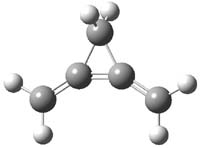
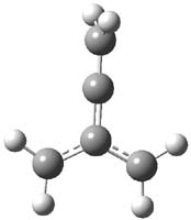
Earlier this year, Barboiu made the astonishing claim of the x-ray characterization of 1,3-dimethylcyclobutadiene, brought about by the photolysis of 4,6-dimethyl-α-pyrone encapsulated in a guanidinium-sulfonate-calixarene crystal (Reaction 1).1 I had not blogged on this paper because Henry Rzepa did a quite thorough analysis of it in this blog post. Now, a couple of rebuttals have appeared and it is time to examine this study.

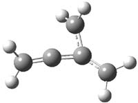
Alabugin calls in question whether the reaction has in fact proceeded beyond 2.2 They note that in the x-ray crystal structure, the distance between a carbon of the purported cyclobutadiene ring and the carbon of CO2 is only 1.50 and 1.61 Å. Barboiu called this a “strong van der Waals contact”, but this is a distance much more attributable to a covalent bond. In fact, the shorter distance is in fact shorter than some of the other C-C distances in the structure that Barboiu calls covalent! Perhaps more bizarre is that the putative CO2 fragment is highly bent: 119.9&;deg;, a value inconsistent with CO2 but perfectly ordinary for an sp2 carbon.  In fact, B3LYP/6-31G** computations suggest that bending CO2 this much costs about 75 kcal mol-1 – and tack on another 7 kcal mol-1 to make the two C-O distances unequal (as found in the x-ray structure!). Thus, Alabugin suggests that only 2 has been formed, and notes that the cleavage to 3 would likely require light of much higher energy that that used in the Barboiu experiment.

Scheschkewitz argues that the x-ray data can be better interpreted as suggesting only the Dewar β-lactone 2 is present, though in its two enantiomeric forms.3 There is no evidence, he suggests of any cyclobutadiene component at all.

It should be noted that Barboiu stands4 by his original work and original assignment, claiming that these types of x-ray experiments are quite difficult and large error bars in atom positions are inherent to the study.

Henry Rzepa has blogged again on this controversy and has a paper coming out on this soon. I shall update when it appears. Henry notes in one of the comments to his blog that a TD-DFT computations does show that the Dewar β-lactone 2 is transparent from 320-500nm.

References

(1) Legrand, Y.-M.; van der Lee, A.; Barboiu, M., "Single-Crystal X-ray Structure of 1,3-Dimethylcyclobutadiene by Confinement in a Crystalline Matrix," Science 2010, 329, 299-302, DOI: 10.1126/science.1188002.

(2) Alabugin, I. V.; Gold, B.; Shatruk, M.; Kovnir, K., "Comment on "Single-Crystal X-ray Structure of 1,3-Dimethylcyclobutadiene by Confinement in a Crystalline Matrix"," Science, 330, 1047, DOI: 10.1126/science.1196188.

(3) Scheschkewitz, D., "Comment on "Single-Crystal X-ray Structure of 1,3-Dimethylcyclobutadiene by Confinement in a Crystalline Matrix"," Science 2010, 330, 1047, DOI: 10.1126/science.1195752.

(4) Legrand, Y.-M.; van der Lee, A.; Barboiu, M., "Response to Comments on "Single-Crystal X-ray Structure of 1,3-Dimethylcyclobutadiene by Confinement in a Crystalline Matrix"," Science, 330, 1047, DOI: 10.1126/science.1195846.

InChIs

1: InChI=1/C7H8O2/c1-5-3-6(2)9-7(8)4-5/h3-4H,1-2H3
InChIKey=IXYLIUKQQQXXON-UHFFFAOYA

2: InChI=1/C7H8O2/c1-4-3-7(2)5(4)6(8)9-7/h3,5H,1-2H3
InChIKey=GLYAMHMFKKLRAL-UHFFFAOYAT

3: InChI=1/C6H8/c1-5-3-6(2)4-5/h3-4H,1-2H3
InChIKey=ADQGKIKNUMJFSL-UHFFFAOYAU

Aromaticity &cyclobutadiene Steven Bachrach 22 Nov 2010 5 Comments

« Previous PageNext Page »