Archive for the 'Molecules' Category

Structure of dihydroxycarbene

Dihdroxycarbene was the subject of a post a few years ago relating to how this carbene does not undergo tunneling,1 while related hydroxycarbene do undergo a tunneling rearrangement.

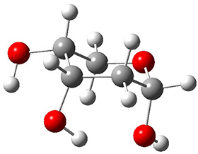
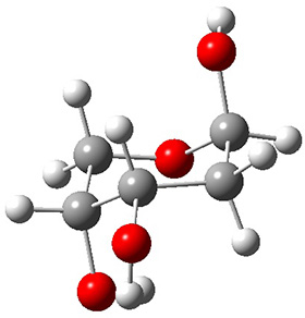
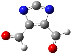
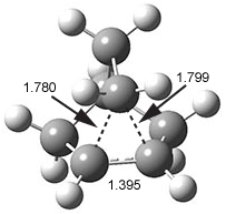
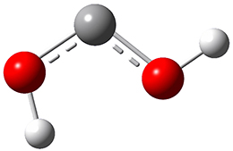
Now we have a gas-phase microwave determination of the trans,cis isomer of dihydroxycarbene.2 The computed CCSD(T)/cc-pCVQZ structure is shown in Figure 1. What is truly remarkable here is the amazing agreement between the experimental and computed structure – as seen in Table 1.The bond distance are in agreement within 0.001 Å and the bond angles agree within 0.3°! Just further evidence of the quality one can expect from high-level computations. And computing this structure was certainly far easier than the experiments!

Figure 1. CCSD(T)/cc-pCVQZ optimized geometry of dihydroxycarbene.

Table 1. Experimental and computed (CCSD(T)/cc-pCVQZ) geometric parameters of dihydroxycarbene.a


Expt.

Comp.

C-O

1.335

1.336

C-O

1.309

1.309

O-Htrans

0.961

0.960

O-Hcis

0.976

0.975

O-C-O

107.30

107.25

C-O-H­­trans

106.8

106.8

C-O-H­­cis

110.7

110.4


aDistances in Å and angles in deg.

References

(1) Schreiner, P. R.; Reisenauer, H. P. "Spectroscopic Identification of Dihydroxycarbene," Angew. Chem. Int. Ed. 2008, 47, 7071-7074, DOI: 10.1002/anie.200802105.

(2) Womack, C. C.; Crabtree, K. N.; McCaslin, L.; Martinez, O.; Field, R. W.; Stanton, J. F.; McCarthy, M. C. "Gas-Phase Structure Determination of Dihydroxycarbene, One of the Smallest Stable Singlet Carbenes," Angew. Chem. Int. Ed. 2014, 53, 4089-4092, DOI: 10.1002/anie.201311082.

InChIs

Dihydroxycarbene: InChI=1S/CH2O2/c2-1-3/h2-3H
InChIKey=VZOMUUKAVRPMBY-UHFFFAOYSA-N

carbenes Steven Bachrach 09 Jun 2014 No Comments

Polytwistane!

Twistane 1 is a more strained isomer of adamantane 2. The structure of 1 is shown in Figure 1.

1

Figure 1. B3LYP/6-31G(d) optimized structure of 1.

Adamantane is the core structure of diamond, which can be made by appending isobutene groups onto adamantane. In an analogous fashion, twistane can be extended in a linear way by appending ethano groups in a 1,4-bridge. Allen, Schreiner, Trauner and co-workers have examined this “polytwistane” using computational techniques.1 They examined a (CH)236 core fragment of polytwistane, with the dangling valences at the edges filled by appending hydrogens, giving a C236H242 compound. This compound was optimized at B3LYP/6-31G(d) and shown in Figure 2a. (Note that I have zoomed in on the structure, but by activating Jmol – click on the figure – you can view the entire compound.) A fascinating feature of polytwistane is its helical structure, which can be readily seen in Figure 2b. A view down the length of this compound, Figure 2c, displays the opening of this helical cylinder; this is a carbon nanotube with an inner diameter of 2.6 Å.

(a)


(b)


(c)

Figure 2. B3LYP/6-31G(d) structure of the C236H242 twistane. (a) A zoomed in look at the structure. This structure links to the Jmol applet allowing interactive viewing of the molecule – you should try this! (b) a side view clearly showing its helical nature. (c) A view down the twistane showing the nanotube structure.

Though the molecule looks quite symmetric, each carbon is involved in three C-C bonds, and each is of slightly different length. The authors go through considerable detail about addressing the symmetry and proper helical coordinates of polytwistane. They also estimate a strain energy of about 1.6 kcal mol-1 per CH unit. This modest strain, they believe, suggests that polytwistanes might be reasonable synthetic targets.

References

(1) Barua, S. R.; Quanz, H.; Olbrich, M.; Schreiner, P. R.; Trauner, D.; Allen, W. D. "Polytwistane," Chem. Eur. J. 2014, 20, 1638-1645, DOI: 10.1002/chem.201303081.

InChIs

1: InChI=1S/C10H16/c1-2-8-6-9-3-4-10(8)5-7(1)9/h7-10H,1-6H2
InChIKey=AEVSQVUUXPSWPL-UHFFFAOYSA-N

2: InChI=1S/C10H16/c1-7-2-9-4-8(1)5-10(3-7)6-9/h7-10H,1-6H2
InChIKey=ORILYTVJVMAKLC-UHFFFAOYSA-N

Schreiner &twistane Steven Bachrach 20 May 2014 3 Comments

Corannulene bowl inversion inside a host

The concept of complementarity between enzyme and substrate, especially the transition state for reactions at the substrate, is a key element of Pauling’s model for enzymatic activity. Koshland’s “induced fit” modification suggests that the enzyme might change its structure during the binding process to either destabilize the reactant or help stabilize the TS. These concepts are now tested in a very nice model by Stoddart, Siegel and coworkers.1

Stoddart recently reported the host compound ExBox4+ 1 and demonstrated that it binds planar polycyclic aromatic hydrocarbons.2 (I subsequently reported DFT computations on this binding.) The twist in this new paper is the binding of corranulene 2 inside ExBox4+ 1. Corranulene is bowl-shaped, with a bowl inversion barrier of 11.5 kcal mol-1 (10.92 kcal mol-1 at B97D/Def2-TZVPP).

The corranulene bowl is too big to fit directly into 1 without some distortions. The x-ray structure of the complex of 1 with 2 inside shows the width of 1 expanding by 0.87 Å and the bowl depth of 2 decreasing by 0.03 Å. The B97D/Def2-TZVPP optimized geometry of this complex (shown in Figure 1) shows similar distortions – the width of 1 increases by 0.37 Å (gas) or 0.29 Å (acetone solution), while the bowl depth of 2 decreases by 0.03 Å (gas) or 0.02 Å (solution).

ground state

transition state

Figure 1. B97D/Def2-TZVPP optimized geometries of the complex of 2 inside 1 (a) ground state and (b) transition state.

The calculated structure of the bowl inversion transition state of 2 inside of 1 is shown in Figure 1. 2 is planar at the TS. The experimental inversion barrier (determined by variable temperature NMR line shift analysis) is 7.88 kcal mol-1, while the calculated barrier is 8.77 kcal mol-1. The reduction in the bowl inversion barrier of 2 inside of 1 is therefore about 2.5 kcal mol-1. The authors argue that this barrier reduction can be attributed to about 0.5 kcal mol-1 of destabilization of the ground state of 2 along with 2 kcal mol-1 of stabilization of the transition state afforded by the host. This study thus confirms the notions of a host reducing a barrier (through both transition state stabilization and ground state destabilization) and induced fit.

References

(1) Juríček, M.; Strutt, N. L.; Barnes, J. C.; Butterfield, A. M.; Dale, E. J.; Baldridge, K. K.; Stoddart, J. F.; Siegel, J. S. "Induced-fit catalysis of corannulene bowl-to-bowl inversion," Nat. Chem. 2014, 6, 222-228, DOI: 10.1038/nchem.1842.

(2) Barnes, J. C.; Juríček, M.; Strutt, N. L.; Frasconi, M.; Sampath, S.; Giesener, M. A.; McGrier, P. L.; Bruns, C. J.; Stern, C. L.; Sarjeant, A. A.; Stoddart, J. F. "ExBox: A Polycyclic Aromatic Hydrocarbon Scavenger," J. Am. Chem. Soc. 2012, 135, 183-192, DOI: 10.1021/ja307360n.

(3) Bachrach, S. M. "DFT Study of the ExBox·Aromatic Hydrocarbon Host–Guest Complex," J. Phys. Chem. A 2013, 117, 8484-8491, DOI: 10.1021/jp406823t.

InChIs

1: InChI=1S/C48H40N4/c1-2-38-4-3-37(1)33-49-25-17-45(18-26-49)41-9-11-43(12-10-41)47-21-29-51(30-22-47)35-39-5-7-40(8-6-39)36-52-31-23-48(24-32-52)44-15-13-42(14-16-44)46-19-27-50(34-38)28-20-46/h1-32H,33-36H2/q+4
InChIKey=ZMELWAYDWQWNOQ-UHFFFAOYSA-N

2: InChI=1S/C20H10/c1-2-12-5-6-14-9-10-15-8-7-13-4-3-11(1)16-17(12)19(14)20(15)18(13)16/h1-10H
InChIKey=VXRUJZQPKRBJKH-UHFFFAOYSA-N

annulenes &host-guest Steven Bachrach 20 Mar 2014 No Comments

The complex PES for sesquiterpene formation

Hong and Tantillo1 report a real tour de force computational study of multiple pathways along the routes towards synthesis of a variety of sesquiterpenes. The starting point is the bisabolyl cation 1, and a variety of rearrangements, cyclizations, proton and hydride transfers are examined to convert it into such disparate products as barbatene 2, widdradiene 3, and champinene 4. The pathways are explored at mPW1PW91/6-31+G(d,p)//B3LYP/6-31+G(d,p). Some new pathways are proposed but the main points are the sheer complexity of the C15H25+ potential energy surface and the interconnections between potential intermediates.

References

(1) Hong, Y. J.; Tantillo, D. J. "Branching Out from the Bisabolyl Cation. Unifying Mechanistic Pathways to Barbatene, Bazzanene, Chamigrene, Chamipinene, Cumacrene, Cuprenene, Dunniene, Isobazzanene, Iso-γ-bisabolene, Isochamigrene, Laurene, Microbiotene, Sesquithujene, Sesquisabinene, Thujopsene, Trichodiene, and Widdradiene Sesquiterpenes," J. Am. Chem. Soc. 2014, 136, 2450-2463, DOI: 10.1021/ja4106489.

InChIs

1: InChI=1S/C15H25/c1-12(2)6-5-7-14(4)15-10-8-13(3)9-11-15/h6,8,15H,5,7,9-11H2,1-4H3/q+1
InChIKey=YKHXORRQMGBNFI-UHFFFAOYSA-N

2: InChI=1S/C15H24/c1-11-6-9-13(2)10-12(11)14(3)7-5-8-15(13,14)4/h6,12H,5,7-10H2,1-4H3/t12-,13-,14+,15-/m0/s1
InChIKey=RMKQBFUAKZOVPQ-XQLPTFJDSA-N

3: InChI=1S/C15H24/c1-12-6-7-13-14(2,3)9-5-10-15(13,4)11-8-12/h6-7H,5,8-11H2,1-4H3/t15-/m0/s1
InChIKey=SJUIWFYSWDVOEQ-HNNXBMFYSA-N

4: InChI=1S/C15H24/c1-11-6-9-15-10-12(11)14(15,4)8-5-7-13(15,2)3/h6,12H,5,7-10H2,1-4H3/t12-,14-,15-/m1/s1
InChIKey=XRDHEPAYTVHOPC-BPLDGKMQSA-N

non-classical &terpenes Steven Bachrach 13 Mar 2014 2 Comments

Monosaccharide PES

The conformational space of monosaccharides is amazingly complex. If we consider just the pyranose form, the ring can in principal exist as a chair, a half-chair, skew (or twist boat) and boat form, for a total of 38 puckering configurations. Layer on top of this the axial and equatorial positions of the hydroxyl and methylhydroxyl groups, and then the rotamers of these substituents, and one is faced with a dauntingly vast space. It is just this space that Beckham and co-workers1 take on for α- and β-glucose, β-xylose, β-mannose and β-acetylglucosamine.

For each sugar, and for each of the 38 puckering configurations, full rotamer scans for each of the substituents led to 27,702 conformations of each of the four monosaccharides, and 36,936 conformations of β-acetylglucosamine. This totals to over 123,000 geometry optimizations that were carried out at M06-2x/6-31G(d). Then taking the structures within 5 kcal mol-1 of the lowest energy structure for ­each pucker, they reoptimized at M06-2X/6-31+G(d,p). Pruning once again those structures that were above 5 kcal mol-1 of the minimum, they performed CCSD(T)/6-311+G(d,p)//B3LYP/6-311+G(2df,p) computations. What a tour de force!

The results of these conformational space surveys are not terribly exciting. The substituents do make a difference in dictating the most and least favorable structures and the activation barriers for interconversion of ring forms.

These PESs will be quite useful in understanding carbohydrate conformations and the role these may play in their chemistry. But the point of bringing this paper to your attention is the tremendously complex, detailed PES that is uncovered, representing the scale of what can be done with modern computers and modern algorithms.

References

(1) Mayes, H. B.; Broadbelt, L. J.; Beckham, G. T. "How Sugars Pucker: Electronic Structure Calculations Map the Kinetic Landscape of Five Biologically Paramount Monosaccharides and Their Implications for Enzymatic Catalysis," Journal of the American Chemical Society 2013, 136, 1008-1022, DOI: 10.1021/ja410264d.

InChIs

α-glucose: InChI=1S/C6H12O6/c7-1-2-3(8)4(9)5(10)6(11)12-2/h2-11H,1H2/t2-,3-,4+,5-,6+/m1/s1
InChIKey: WQZGKKKJIJFFOK-DVKNGEFBSA-N

β-glucose: InChI=1S/C6H12O6/c7-1-2-3(8)4(9)5(10)6(11)12-2/h2-11H,1H2/t2-,3-,4+,5-,6-/m1/s1
InChIKey=WQZGKKKJIJFFOK-VFUOTHLCSA-N

β-xylose: InChI=1S/C5H10O5/c6-2-1-10-5(9)4(8)3(2)7/h2-9H,1H2/t2-,3+,4-,5-/m1/s1
InChIKey=SRBFZHDQGSBBOR-KKQCNMDGSA-N

β-mannose: InChI=1S/C6H12O6/c7-1-2-3(8)4(9)5(10)6(11)12-2/h2-11H,1H2/t2-,3-,4+,5+,6-/m1/s1
InChIKey=WQZGKKKJIJFFOK-RWOPYEJCSA-N

β-acetylglucosamine: InChI=1S/C8H15NO6/c1-3(11)9-5-7(13)6(12)4(2-10)15-8(5)14/h4-8,10,12-14H,2H2,1H3,(H,9,11)/t4-,5-,6-,7-,8-/m1/s1
InChIKey=OVRNDRQMDRJTHS-FMDGEEDCSA-N

sugars Steven Bachrach 18 Feb 2014 1 Comment

Gas phase structure of 2-deoxyribose

2-deoxyribose 1 is undoubtedly one of the most important sugars as it is incorporated into the backbone of DNA. The conformational landscape of 1 is complicated: it can exist as an open chain, as a five-member ring (furanose), or a six-member ring (pyranose), and intramolecular hydrogen bonding can occur. This internal hydrogen bonding is in competition with hydrogen bonding to water in aqueous solution. Unraveling all this is of great interest in predicting structures of this and a whole host of sugar and sugar containing-molecules.


1

In order to get a firm starting point, the gas phase structures of the low energy conformers of 1 would constitute a great set of structures to use as a benchmark for gauging force fields and computational methods. Cocinero and Alonso1 have performed a laser ablation molecular beam Fourier transform microwave (LA-MB-FTMW) experiment (see these posts for other studies using this technique) on 1 and identified the experimental conformations by comparison to structures obtained at MP2/6-311++G(d,p). Unfortunately the authors do not include these structures in their supporting materials, so I have optimized the low energy conformers of 1 at ωB97X-D/6-31G(d) and they are shown in Figure 1.

1a (0.0)

1b (4.7)

1c (3.3)

1d (5.6)

1e (8.9)

1f (9.4)

Figure 1. ωB97X-D/6-31G(d) optimized structures of the six lowest energy conformers of 1. Relative free energy in kJ mol-1.

The computed spectroscopic parameters were used to identify the structures responsible for the six different ribose conformers observed in the microwave experiment. To give a sense of the agreement between the computed and experimental parameters, I show these values for the two lowest energy conformers in Table 1.

Table 1. MP2/6-311++G(d,p) computed and observed spectroscopic parameters for the two lowest energy conformers of 1.


1a

1c


Expt

Calc

Expt

Calc

A(MHz)

2484.4138

2492

2437.8239

2447

B (MHz)

1517.7653

1533

1510.7283

1527

C (MHz)

1238.9958

1250

1144.9804

1158

ΔG (kJ mol-1)


0.0


3.3

This is yet another excellent example of the symbiotic relationship between experiment and computation in structure identification.

References

(1) Peña, I.; Cocinero, E. J.; Cabezas, C.; Lesarri, A.; Mata, S.; Écija, P.; Daly, A. M.; Cimas, Á.; Bermúdez, C.; Basterretxea, F. J.; Blanco, S.; Fernández, J. A.; López, J. C.; Castaño, F.; Alonso, J. L. "Six Pyranoside Forms of Free 2-Deoxy-D-ribose," Angew. Chem. Int. Ed. 2013, 52, 11840-11845, DOI: 10.1002/anie.201305589.

InChIs

1a: InChI=1S/C5H10O4/c6-3-1-5(8)9-2-4(3)7/h3-8H,1-2H2/t3-,4+,5-/m0/s1
InChIKey=ZVQAVWAHRUNNPG-LMVFSUKVSA-N

MP &sugars Steven Bachrach 16 Dec 2013 No Comments

Predicting reactive C-C bonds

Can one identify a labile bond in a molecule without computing activation barriers? Markopoulos and Grunenberg suggest that examination of the bond length and its associated relaxed force constant might provide some guidance.1

The relaxed force constant comes from identifying the force constant for some coordinate while allowing for other coordinates to relax. Badger’s rule relates the (normal) force constant to bond distance (k = a/(reqd)3). For a series of 36 molecules, covering 71 C-C single bonds, Badger’s rule fits the data well, except for a set of molecules which undergo rapid Cope rearrangements (like bullvalene and semibullvalene). For these molecules, the relaxed force constants are much lower than Badger’s rule predicts, and indicates a weakened bond. This gives rise to their low activation barriers.

Another example is provided with the highly strained polycyclic hydrocarbon 1. This compound is predicted (B3LYP/6-31G(d)) to undergo a [1,2]-shift to give the carbene 2 (see Figure 1), and this is extremely exothermic: -105.7 kcal mol-1, reflecting the enormous strain of 1. The barrier, through TS1 (Figure 1), is only 6.7 kcal mol-1. This rearrangement was predicted by examining the relaxed force constants which identified a very weak bond, despite a short bond distance of 1.404 Å. It is unlikely that without this guidance, one would have predicted that this short bond is likely to rupture and produce this particular product.

1

2

TS1

Figure 1. B3LYP/6-31G(d) optimized structures of 1, 2, and TS1.

References

(1) Markopoulos, G.; Grunenberg, J. "Predicting Kinetically Unstable C-C Bonds from the Ground-State Properties of a Molecule," Angew. Chem. Int. Ed. 2013, 52, 10648-10651, DOI: 10.1002/anie.20130382.

InChIs

1: InChI=1S/C14H12/c1-2-8-11-5-3-9-7(1)10(9)4-6-12(8,11)14(8,11)13(7,9)10/h1-6H2
InChIKey=LNBZAENQMFDBJW-UHFFFAOYSA-N

2: InChI=1S/C14H12/c1-3-11-12-4-2-9-7-8(1,9)10(9)5-6-13(11,12)14(10,11)12/h1-6H2
InChIKey=UKVODHRLGFPZPT-UHFFFAOYSA-N

carbenes Steven Bachrach 25 Nov 2013 3 Comments

Unusual carbene ground states

The singlet and triplet carbene is the topic of Chapter 4, especially sections 1 and 2. The ground state of methylene is the triplet, with one electron in the σ-orbital and one electron in the π-orbital, with the spins aligned. The lowest singlet state places the pair of electrons in the σ-orbital, and this state is about 9 kcal mol-1 higher in energy than the triplet. The next lowest singlet state has one electron in each of the σ- and π-orbitals, with the spins aligned. The singlet state with both electrons in the π-orbital is the highest of these four states, some 60 kcal mol-1 above the ground state triplet.

Hoffmann and Borden now pose the question “Can the doubly occupied π carbene (1A10π2) be the ground state with appropriate substitution?” The answer they find is yes!1

The trick is to find a combination of substituents that will raise the energy of the σ-orbital and lower the energy of the π-orbital. The latter effect can be enhanced if the π-orbital can be a part of an aromatic (6e) ring.

Two of the best possibilities for identifying a ground state 1A10π2 carbene are 1 and 2. The CASSCF/6-31G(d) optimized geometries of these two are shown in Figure 1. In 1, the nitrogen lone pairs act to destabilize the σ-orbital, while the aldehyde group acts as a withdrawing group to stabilize the π-orbital. The result is that the 1A10π6 state of 1 is predicted to be about 8 kcal mol-1 more stable than the triplet state, as per CASPT2 and CCSD(T) computations.


1


2

An ever greater effect is predicted for 2. Here the nitrogen lone pairs adjacent to the carbene act to destabilize the σ-orbital. The empty π-orbital on B lowers the energy of the carbene π-orbital by making it part of the 6-electron aromatic ring. The 1A10π6 state of 2 is predicted to be about 25 kcal mol-1 more stable than its triplet state!

1

2

Figure 1. CASSCF/6-31G(d) optimized geometries of the 1A10π6 states of 1 and 2.

References

(1) Chen, B.; Rogachev, A. Y.; Hrovat, D. A.; Hoffmann, R.; Borden, W. T. "How to
Make the σ0π2 Singlet the Ground State of Carbenes," J. Am. Chem. Soc. 2013, 135, 13954-13964, DOI: 10.1021/ja407116e.

InChIs

1: InChI=1S/C5H2N2O2/c8-1-4-5(2-9)7-3-6-4/h1-2H
InChIKey=QDSVROXEBBWIOO-UHFFFAOYSA-N

2: InChI=1S/C3H3BN2/c1-4-2-6-3-5-1/h1-2,4H
InChIKey=MQJXDZBYOSOLST-UHFFFAOYSA-N

Borden &carbenes Steven Bachrach 14 Oct 2013 4 Comments

The x-ray structure of norbornyl cation

A long sought-after data point critical to the non-classical cation story has finally been obtained. The elusive x-ray crystal structure of a norbornyl cation was finally solved.1 The [C7H11]+[Al2Br7] salt was crystallized in CH2Br2 at low temperature (40 K). This low temperature was needed to prohibit rotation of the norbornyl cation within the crystal (the cation is near spherical and so subject to relatively easy rotation within the crystal matrix) and hydride scrambling among the three carbons (C1, C2, and C6) involved in the non-classical cation structure.

The authors report a number of different structures, all very similar, depending on slight differences in the crystals used. However, the important features are consistent with all of the structures. The cation is definitely of the non-classical type (see Figure 1) with the basal C1-C2 bond length of 1.39 Å similar that in benzene and long non-classical C1-C6 and C2-C6 distances of 1.80 Å. These distances match very well with the MP2(FC)/def2-QZVPP optimized distances of 1.393 and 1.825 Å, respectively.

Figure 1. X-ray structure of norbornyl cation.

References

(1) Scholz, F.; Himmel, D.; Heinemann, F. W.; Schleyer, P. v. R.; Meyer, K.; Krossing, I. "Crystal Structure Determination of the Nonclassical 2-Norbornyl Cation," Science 2013, 341, 62-64, DOI: 10.1126/science.1238849.

non-classical &norbornyl cation &Schleyer Steven Bachrach 16 Sep 2013 No Comments

Nonamethylcyclopentyl cation

The nine methyl groups of nonamethylcyclopentyl cation 1 all interconvert with a barrier of 7 kcal mol-1. However, at low temperature only partial scrambling occurs: there are two sets of methyl groups, one containing five groups and the other containing four methyl groups. The barrier for this scrambling is only 2.5 kcal mol-1. While this behavior was found more than 20 years ago, Tantillo and Schleyer1 only now have offered a complete explanation.


1

The ground state structure of 1 is shown in Figure 1 and has C1 symmetry. The two pseudo-axial methyl groups adjacent to the cationic center show evidence of hyperconjugation: long C-C bonds and Me-C-C+ angles of 100°.

The transition state TS1¸also in Figure 1, is of Cs symmetry. This transition state leads to interchange of the pseudo-axial methyls, and interchange of the pseudo-equatorial methyls, but no exchange between the members of these two groups. The M06-2x/6-31+G(d,p) and mPW1PW91/6-31+G(d,p) estimate of this barrier is 1.5 and 2.5 kcal mol-1, respectively. This agrees well with the experiment.

1

TS1

TS2

Figure 1. B3LYP/6-3+G(d,p) optimized geometries.

A second transition state TS2 was found and it corresponds with a twisting motion that interconverts an axial methyl with an equatorial methyl. This TS has Cs symmetry (shown in Figure 1) and the eclipsing interaction give rise to a larger barrier: 7.3 (M06-2x/6-31+G(d,p)) and 6.7 kcal mol-1 (mPW1PW91/6-31+G(d,p)). So twisting through TS2 and scrambling through TS1 allows for complete exchange of all 9 methyl groups.

An interesting point also made by these authors is that these three structures represent the continuum of cationic structure: a classical (localized) cation in TS2, a bridged structure in TS1 and hyperconjugated cation in 1.

References

(1) Tantillo, D. J.; Schleyer, P. v. R. “Nonamethylcyclopentyl Cation Rearrangement Mysteries Solved,” Org. Lett. 2013, 15, 1725-1727, DOI: 10.1021/ol4005189.

InChIs

1: InChI=1S/C14H27/c1-10-11(2,3)13(6,7)14(8,9)12(10,4)5/h1-9H3/q+1
InChIKey=WUGVCUSQGLXERW-UHFFFAOYSA-N

non-classical &Schleyer Steven Bachrach 23 Jul 2013 4 Comments

« Previous PageNext Page »