Structure of conicasterol F

NMR Steven Bachrach 01 May 2012 No Comments

Here is an interesting twist on using computations in conjunction with experimental NMR to solve for molecular structure. I have blogged a number of times on comparing computed chemical shifts with experimental values to identify structure, and also on using the comparison of computed and experimental coupling constants to accomplish this purpose.

Butts and Bifulco were interested in the structure of conicasterol F 1 and opted to make two sets of comparison.1 The first uses the traditional approach of comparing the computed and experimental 13C chemical shifts. The second comparison uses the distances between protons, coming from the optimized structure and the rotating-frame nuclear Overhauser effect (ROE).

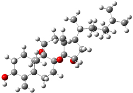
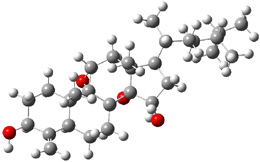
Standard analysis of the NMR spectra of 1 allowed for the determination of all of the stereochemistry except for the epoxy ring at C8 and C14. The possible options are shown as 1a and 1b. The optimized geometries (MPW1PW91/6-31G) of these two diastereomers are shown in Figure 1.


1a


1b

1a

1b

Figure 1. Optimized geometries of 1a and 1b.

Comparison of 15 distances between protons determined by the ROE experiment and by computation led to a mean absolute error of 7.8% for 1a and 3.0% for 1b, suggesting that the latter is the correct structure. Similar comparison was then made between the experimental chemical shifts of 12 of the carbon atoms with the computed values of the two isomers. The mean absolute error in the chemical shifts of 1a is 3.7ppm, but only 0.8 ppm for 1b. Both methods give the same conclusion: conicasterol F has structure 1b.

References

(1) Chini, M. G.; Jones, C. R.; Zampella, A.; D’Auria, M. V.; Renga, B.; Fiorucci, S.; Butts, C. P.; Bifulco, G., "Quantitative NMR-Derived Interproton Distances Combined with Quantum Mechanical Calculations of 13C Chemical Shifts in the Stereochemical Determination of Conicasterol F, a Nuclear Receptor Ligand from Theonella swinhoei," J. Org. Chem., 2012, 77, 1489-1496, DOI: 10.1021/jo2023763.

InChIs

1b: InChI=1/C29H46O4/c1-16(2)17(3)8-9-18(4)21-14-23(31)28-26(21,7)15-24-29(32-24)25(6)12-11-22(30)19(5)20(25)10-13-27(28,29)33-28/h16-18,20-24,30-31H,5,8-15H2,1-4,6-7H3/t17-,18-,20+,21-,22+,23+,24-,25+,26-,27+,28+,29+/m1/s1
InChIKey=XTWHHLLDFQALAM-XAIGNWORBC

Enzymatic catalysis of ladder ether formation

Enzyme Steven Bachrach 24 Apr 2012 1 Comment

Biosynthesis of ladder polyethers is the topic of a very nice experimental/computational study by Chen and Houk.1 The x-ray structure of the enzyme that catalyzes the nucleophilic attack on epoxides to create the 6-member ring ether was determined, but the geometry did not completely indicate the mechanism.

Gas phase computations of the 5-exo-tet and 6-endo-tet ring openings of 1 were examined for both the acid and base catalyzed routes at B2PLYP/6-311++G(d,p)//B2PLYP/6-31G(d).
The results are summarized in Figure 1. Basically, as expected by Baldwin’s rules, the closure to the tetrahydrofuran (5-exo-tet) is favored under both catalyzed conditions. However, the preference is small under base conditions, with the difference in the free energy of activation of only 1.2 kcal mol-1.

Figure 1. Gas phase energies (kcal mol-1) for the acid an base catalyzed reactions of 1 to 2 or 3.

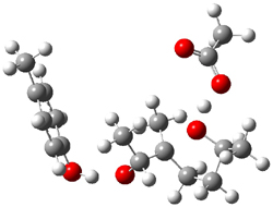
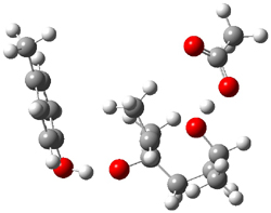
The enzyme Lsd19B produces just the analogue of 3. So, the two regioisomeric TSs were reoptimized with an aspartic acid group and a tyrosine group in the positions they occupy in the active site of the enzyme Lsd19b. The two resulting transition states, evaluated at B2LYP/6-311++G(d,p)//MO6-2x/6-31G(d), are shown in Figure 2. The activation energy for the 6-endo-tet reaction is 18.0 kcal mol-1, 2.5 kcal mol-1 lower than for the 5-exo-tet route. This energy difference would give rise to a 100:1 selectivity for the tetrahydropyran product, in accord with experiment. The enzyme preorganizes for and favors the base catalyzed path that leads to 3.

5-exo-tet TS model
ΔG‡ = 20.5

6-endo-tet TS model
ΔG‡ = 18.0

Figure 2. Transition state models of the active site. Activation energies in kcal mol-1.

References

(1) Hotta, K.; Chen, X.; Paton, R. S.; Minami, A.; Li, H.; Swaminathan, K.; Mathews, I. I.; Watanabe, K.; Oikawa, H.; Houk, K. N.; Kim, C.-Y., "Enzymatic catalysis of anti-Baldwin ring closure in polyether biosynthesis," Nature, 2012, 483, 355-358, DOI: 10.1038/nature10865.

InChIs

1: InChI=1/C8H16O2/c1-6(9)4-5-8(3)7(2)10-8/h6-7,9H,4-5H2,1-3H3/t6-,7?,8+/m0/s1
InChIKey=YEAGKBPXLVGCPK-YPVSKDHRBB

2: InChI=1/C8H16O2/c1-6-4-5-8(3,10-6)7(2)9/h6-7,9H,4-5H2,1-3H3/t6-,7-,8-/m0/s1
InChIKey=RRDMOYCGUXNJSL-FXQIFTODBG

3: InChI=1/C8H16O2/c1-6-4-5-8(3,9)7(2)10-6/h6-7,9H,4-5H2,1-3H3/t6-,7-,8+/m0/s1
InChIKey=WWLWIBPPUNCAQU-BIIVOSGPBQ

Fluoresence of encapsulated stilbene

stilbene Steven Bachrach 10 Apr 2012 No Comments

Petsalakis and Rebek have explored the fluorescence of stilbene inside a couple of different kinds of capsules.1 trans-Stilbene exhibits weak fluorescence in solution, but when placed inside a small capsule, the fluorescence disappears almost entirely, while in a large capsule, the fluorescence returns to normal. They examined stilbene inside two different capsules using a variety of DFT and ONIOM techniques.

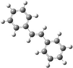
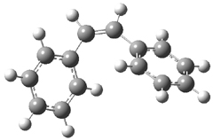
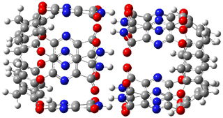
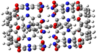
The optimized geometries of trans- and cis-stilbene optimized at CAM-B3LYP/6-31G(d,p) are displayed in Figure 1. As expected, the trans conformer is planar and the cis conformer is twisted to avoid clashes between the phenyl rings. The optimized structure of the 1.1 capsule is also shown in Figure 1. All of these structures are fairly insensitive to computational method. (They have also looked at an even larger capsule, but I have omitted displaying its structure here.)

(a)

(b)

(c)

(d)

Figure 1. CAM-B3LYP/6-31G(d,p) optimized structures of (a) trans-stilbene, (b) cis-stilbene , (c) the 1.1 capsule, and (d) trans-stilbene inside the 1.1 capsule.

The structure of trans- stilbene inside the 1.1 capsule is shown in Figure 1. Of particular note is that the stilbene is no longer planar. (This twisting is perhaps better observed by an end-on view, which the reader can obtain by clicking on the picture and then manipulating the full 3-D structure using the Jmol applet.) The different computational methods give slightly different encapsulated structures, and vary a bit in their binding energies, but the twisting of the stilbene is reproduced by each method. Though not shown here, trans-stilbene in the larger capsule is again a nearly planar structure.

The structure of the S1 state of trans-stilbene in the large capsule is the same as for free trans-stilbene. However, the geometry of the S1 state in the smaller 1.1 capsule is twisted and corresponds to the conical intersection geometry.

The absorption spectra and the emission spectra were computed for the free and encapsulated structures. The absorption and emission spectra for free stilbene and stilbene in the larger capsule are nearly identical, corresponding to the experimental observation of similar fluorescence behavior. The absorption spectra of stilbene in the 1.1 capsule has a small blue shift of 8 nm due to the twisted geometry. But the major result is that the S1 state of stilbene inside the 1.1 capsule distorts to the conical intersection, allowing for radiationless return to the ground state. This means that there would be no fluorescence, and that is exactly what is observed.

References

(1) Tzeli, D.; Theodorakopoulos, G.; Petsalakis, I. D.; Ajami, D.; Rebek, J., "Conformations and Fluorescence of Encapsulated Stilbene," J. Am. Chem. Soc., 2012, 134, 4346-4354, DOI: 10.1021/ja211164b

InChIs

trans-stilbene: InChI=1/C14H12/c1-3-7-13(8-4-1)11-12-14-9-5-2-6-10-14/h1-12H/b12-11+
InChIKey=PJANXHGTPQOBST-VAWYXSNFBV

cis-stilbene: InChI=1/C14H12/c1-3-7-13(8-4-1)11-12-14-9-5-2-6-10-14/h1-12H/b12-11-
InChIKey=PJANXHGTPQOBST-QXMHVHEDBW

C2 and the quadruple bond

Bond Dissociation Energy Steven Bachrach 03 Apr 2012 1 Comment

Inspired by a blog post of Henry Rzepa (see here) Shaik and co-workers examined the C2 species with an eye towards the nature of the bond between the two carbon atoms.1 Using both a valence bond approach and a full CI approach, they end up at the same place: there is a quadruple bond here!

The argument rests largely on a definition of of an in situ bond energy. For the VB approach, this requires choosing as a reference a non-bonding interaction between the atoms with regards to a pair of electrons. For the CI approach, the bond energy is half the energy of the singlet-triplet gap. So, for C2, the VB/6-31G* estimate of the bond energy of the putative fourth bond is 14.3 kcal mol-1. For the full CI/6-31G* computations of the singlet-triplet gap, the bond energy estimate is 14.8 kcal mol-1, and using the experimental value of the gap, the estimate is 13.2 kcal mol-1. Not a strong bond, but certainly meaningful!

In the VB approach, the fourth bond is a weighted sum of the antibonding 2σu and bonding 3σg orbitals – a combination that gives rise to small constructive overlap between the two C atoms. In the CI model, the wavefunction is dominated by the first two configurations; the first configuration, with a coefficient of C0=0.828 has 2σu doubly occupied and the second coefficient, with CD=0.324, has the 3σg orbital doubly occupied. Considering that 3σg is a bonding orbital, the significant contribution of this configuration gives rise to the fourth bond.

References

(1) Shaik, S.; Danovich, D.; Wu, W.; Su, P.; Rzepa, H. S.; Hiberty, P. C., "Quadruple bonding in C2 and analogous eight-valence electron species," Nat. Chem., 2012, 4, 195-200, DOI: 10.1038/nchem.1263.

InChIs

C2: InChI=1/C2/c1-2
InChIKey=LBVWYGNGGJURHQ-UHFFFAOYAE

Nanohoop of linked napthlylene groups

nanohoops Steven Bachrach 27 Mar 2012 No Comments

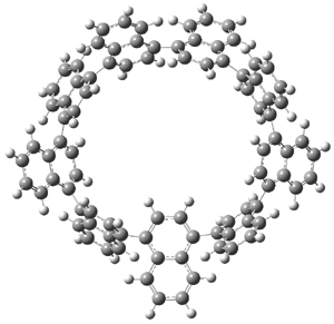
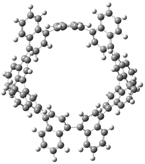
Itami continues to design novel macrocycles containing aromatic rings (see this post). This latest paper reports the synthesis of the first nanohoops containing naphthylenes, namely [9]cyclo-1,4-naphthylene 1.1 Since the macrocycle contains an odd number of naphthylene units, the lowest energy conformation is of C2 symmetry with one of the naphthylene rings in the plane of the macrocycle. (See Figure 1 for the B3LYP/6-31G(d) optimized structure). This conformation gives rise to 27 peaks in the proton NMR, and while the value of the computed chemical shifts differ from the experimental ones by about 0.5 to 1 ppm, their relative ordering is in very nice agreement.

1

2

Figure 1. B3LYP/6-31G(d) optimized geometries of 1 and the racemization transition state 2.

Itami also notes that 1 is chiral and computed the barrier for racemization of 19.9 kcal mol-1¸ through the transition state 2, also shown in Figure 1. This racemization process is compared with the racemization of 1,1’-binaphthyl.

References

(1) Yagi, A.; Segawa, Y.; Itami, K., "Synthesis and Properties of [9]Cyclo-1,4-naphthylene: A π-Extended Carbon Nanoring," J. Am. Chem. Soc. 2012, 134, 2962-2965, DOI: 10.1021/ja300001g

InChIs

1: InChI=1/C90H54/c1-2-20-56-55(19-1)73-37-38-74(56)76-41-42-78(60-24-6-5-23-59(60)76)80-45-46-82(64-28-10-9-27-63(64)80)84-49-50-86(68-32-14-13-31-67(68)84)88-53-54-90(72-36-18-17-35-71(72)88)89-52-51-87(69-33-15-16-34-70(69)89)85-48-47-83(65-29-11-12-30-66(65)85)81-44-43-79(61-25-7-8-26-2(61)81)77-40-39-75(73)57-21-3-4-22-58(57)77/h1-54H/b75-73-,76-74-,79-77-,80-78-,83-81-,84-82-,87-85-,88-86-,90-89-
InChIKey=WUFIQFYLEOBLMY-YNQZQJAJBX

Ethynyl-substituted Cyclobutadiene

cyclobutadiene Steven Bachrach 20 Mar 2012 No Comments

Cyclobutadiene is the prototypical antiaromatic compound. McMahon has examined the
effect of ethynyl substitution on this ring, with a long term eye towards the possibility of these types of species being involved in the synthesis of fullerenes.1

All of the possible ethynyl-substituted cyclobutadiene species (1-7) were optimized at B3LYP/6-31G(d) and CCSD/cc-pVDZ in their singlet and triplet states.

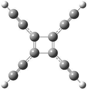
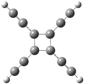
The structures of singlet and triplet 7 are shown in Figure 1. The geometries provided by the two different methods are quite similar. They show a rectangular form for the singlets and a delocalized, nearly square ring for the triplets.

7singlet

7triplet

Figure 1. CCSD/cc-pVDZ optimized structures of singlet and triplet 7.

The computed singlet-triplet gap decreases with each ethynyl substituent. B3LYP, which overestimates the stability of triplets, predicts that 6 and 7 will be ground state triplets, while CCSD predicts a singlet ground state for all 7 species, with the gap decreasing steadily from 11.5 to 8.2 kcal mol-1, a value that is also probably underestimated.

This change in the singlet-triplet gap reflects a stronger stabilizing effect of each ethynyl group to the cycnobutadiene ring for the triplet than for the singlet state. This is seen in the homodesmotic stabilization energies.

Lastly, NICS(1)zz values are positive for all of the singlets and negative for the triplets. The positive values for the singlets reflect their antiaromatic character, also seen in the alternant bond distances around the ring. The NICS values of the singlets decrease with increasing substitution. The negative NICS values of the triplets reflects aromatic character, as seen in the non-alternant distances around the ring. Interestingly, the triplet NICS values decrease with increasing ethynyl substitution, suggesting decreased aromaticity, even though the homodesmotic reactions suggest increasing stabilization with substitution.

References

(1) Esselman, B. J.; McMahon, R. J., "Effects of Ethynyl Substitution on Cyclobutadiene," J. Phys. Chem. A 2012, 116, 483-490, DOI: 10.1021/jp206478q

InChIs

1: InChI=1/C4H4/c1-2-4-3-1/h1-4H
InChIKey=HWEQKSVYKBUIIK-UHFFFAOYAI

2: InChI=1/C6H4/c1-2-6-4-3-5-6/h1,3-5H
InChIKey=XFHXCHFBSJDBGT-UHFFFAOYAG

3: InChI=1/C8H4/c1-3-7-5-6-8(7)4-2/h1-2,5-6H
InChIKey=YSSBCMLQKUIAEP-UHFFFAOYAI

4: InChI=1/C8H4/c1-3-7-5-8(4-2)6-7/h1-2,5-6H
InChIKey=IRAQOGPKMAXTQY-UHFFFAOYAN

5: InChI=1/C8H4/c1-3-7-5-6-8(7)4-2/h1-2,5-6H
InChIKey=YSSBCMLQKUIAEP-UHFFFAOYAI

6: InChI=1/C10H4/c1-4-8-7-9(5-2)10(8)6-3/h1-3,7H
InChIKey=BVCIPPXDFXWTJR-UHFFFAOYAQ

7: InChI=1/C12H4/c1-5-9-10(6-2)12(8-4)11(9)7-3/h1-4H
InChIKey=HEUYILYXFVQGOW-UHFFFAOYAO

Regiolone and isosclerone: enantiomers resolved

Optical Rotation Steven Bachrach 13 Mar 2012 2 Comments

It is striking to me that the absolute configuration of relatively simple compounds remains problematic even today. The structure of two naturally-occurring phytotoxic enantiomers 1, called regiolone and isosclerone, are finally definitively defined using a computational approach.


(R)-1: R = OH, R’ = H
(S)-1: R = H, R’ = OH

Isosclerone is the dextrorotatory isomer, while regiolone is the levorotatory isomer. The question though is which one is R and which one is S? Evidente and co-workers arbitrarily decided to compute the spectral properties of the S isomer.1 They located four low energy conformers at B3LYP/6-31G* and B3LYP/TZVP. (These conformers are not shown here as the authors did not deposit the coordinates. Reviewers and editors – please insist that this computational data be mandatory for publication!) The conformer relative energies, listed in Table 1, are dependent on the method, however, the two lowest energy structures will dominate the population and both will be present to a significant extent, regardless of which energy set is used. The optical rotation [α]D was computed at B3LYP/6-31G*//B3LYP/TZVP, and these too are listed in Table 1. The Boltzmann-weighted [α]D is 21.8. Even though the lowest energy conformer contributes a negative rotation, the much larger positive rotation due to the second-lowest energy conformer, along with the two other conformers, will dominate to dictate the OR value. This suggests that the enantiomers are (S)(+)-1 and (R)(-)-1. Computed ECD spectra confirm this assignment; the computed ECD of the (S) isomer is a near mirror image of the experimental ECD of the (-)-1 compound. Therefore, regiolone is (R)(-)-1 and isosclerone is (S)(+)-1.

Table 1. Relative free energies (kcal mol-1) and [α]D of the conformers of (S)-1.a

conformer

ΔG, 6-31G*

ΔG, TZVP

[α]Db

A 0.43 0.0 -17.50
B 0.0 0.32 67.92
C 1.21 1.03 95.72
D 1.84 1.48 17.72

aAll computations performed with B3LYP. bAt B3LYP/6-31G*//B3LYP/TZVP

References

(1) Evidente, A.; Superchi, S.; Cimmino, A.; Mazzeo, G.; Mugnai, L.; Rubiales, D.; Andolfi, A.; Villegas-Fernández, A. M., "Regiolone and Isosclerone, Two Enantiomeric Phytotoxic Naphthalenone Pentaketides: Computational Assignment of Absolute Configuration and Its Relationship with Phytotoxic Activity," Eur. J. Org. Chem., 2011, 5564-5570, DOI: 10.1002/ejoc.201100941

InChIs

Regiolone: InChI=1/C10H10O3/c11-7-4-5-9(13)10-6(7)2-1-3-8(10)12/h1-3,7,11-12H,4-5H2/t7-/m1/s1
InChIKey=ZXYYTDCENDYKBR-SSDOTTSWBB

Isosclerone: InChI=1/C10H10O3/c11-7-4-5-9(13)10-6(7)2-1-3-8(10)12/h1-3,7,11-12H,4-5H2/t7-/m0/s1
InChIKey=ZXYYTDCENDYKBR-ZETCQYMHBD

More strange dynamics from the Singleton Group

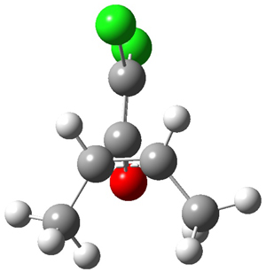
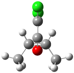
Once again the Singleton group reports experiments and computations that require serious reconsideration of our notions of reaction mechanisms.1 In this paper they examine the reaction of dichloroketene with labeled cis-2-butene. With 13C at the 2 position of 2-butene, two products are observed, 1 and 1’, in a ratio of 1’:1 = 0.993 ± 0.001. This is the opposite what one might have imagined based on the carbonyl carbon acting as an electrophile.

The first interesting item is that B3LYP/6-31+G** fails to predict the proper structure of the transition state. It predicts an asymmetric structure 2, shown in Figure 1, while MPW1k/6-31+G**, M06, and MP2 predict a Cs transition structure 3. The Cs TS is confirmed by a grid search of M06-2x geometries with CCSD(T)/6-311++G88/PCM(CH2Cl2) energies.

2

3

Figure 1. Optimized TSs 2 (B3LYP/6-31+G**) and 3 (MPW1K/6-31+G**).

The PES using proper computational methods is bifurcating past TS 3, falling downhill to product 1 or 1’. Lying on the Cs plane is a second transition state that interconverts 1 and 1’. On such a surface, conventional transition state theory would predict equal amounts of 1 and 1’, i.e. no isotope effect! So they must resort to a trajectory study – which would be impossibly long if not for the trick of making the labeled carbon super-heavy – like 28C,44C, 76C and 140C and then extrapolating back to just ordinary 13C. These trajectories indicate a ratio of 1’:1 of 0.990 in excellent agreement with the experimental value of 0.993.

Interestingly, most trajectories recross the TS, usually by reaching into the region near the second TS. However, the recrossing decreases with increasing isotopic mass, and this leads to the isotope effect. It turns out the vibrational mode 3 breaks the Cs symmetry; movement in one direction along mode 3 has no mass dependence but in the opposite direction, increased mass leads to decreased recrossing – or put in another way, in this direction, increased mass leads more often to product.

But one can understand this reaction from a statistical point of view as well. If one looks at the free energy surface, there is a variational TS near 3, but then there is a second set of variational transition states (one leading to 1 and one to 1’) which are associated with the formation of the second C-C bond. In a sense there is an intermediate past 3 that leads to two entropic barriers, one on a path to 1 and one on the path to 1’. RRKM using this model gives a ratio of 0.992 – again in agreement with experiment! It is as Singleton notes “perplexing”; how do you reconcile the statistical view with the dynamical (trajectory) view? Singleton has no full explanation.

Lastly, they point out that a similar situation occurs in the organocatalyzed Diels-Alder reaction of MacMillan shown below.2 (This reaction is also discussed in a previous post.) Now Singleton finds that the “substituent effects, selectivity, solvent effects, isotope effects and activation parameters” are all dictated by a second variational TS far removed from the conventional electronic TS.

References

(1) Gonzalez-James, O. M.; Kwan, E. E.; Singleton, D. A., "Entropic Intermediates and Hidden Rate-Limiting Steps in Seemingly Concerted Cycloadditions. Observation, Prediction, and Origin of an Isotope Effect on Recrossing," J. Am. Chem. Soc. 2012, 134, 1914-1917, DOI: 10.1021/ja208779k

(2) Ahrendt, K. A.; Borths, C. J.; MacMillan, D. W. C., "New Strategies for Organic Catalysis: The First Highly Enantioselective Organocatalytic Diels-Alder Reaction," J. Am. Chem. Soc. 2000, 122, 4243-4244, DOI: 10.1021/ja000092s.

InChIs

2-butene: InChI=1/C4H8/c1-3-4-2/h3-4H,1-2H3/b4-3-
InChIKey=IAQRGUVFOMOMEM-ARJAWSKDBO

Dichloroketene: InChI=1/C2Cl2O/c3-2(4)1-5
InChIKey=TVWWMKZMZALOFP-UHFFFAOYAY

1 (no isotope): InChI=1/C6H8Cl2O/c1-3-4(2)6(7,8)5(3)9/h3-4H,1-2H3/t3-,4+/m0/s1
InChIKey=BAEYWHUXGUIZSP-IUYQGCFVBH

Welwitindolinones structure

NMR Steven Bachrach 28 Feb 2012 1 Comment

A quick note here on the use of computed NMR to determine stereochemical structure. The Garg group synthesized two “oxidized welwitindolines”, compounds 1 and 2.1 The relative stereochemistry at the C3 position (the carbon with the hydroxy group) was unknown.


1


2

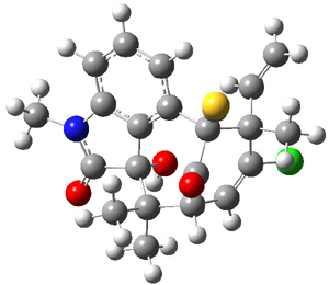
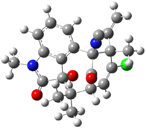
Low energy gas-phase conformers of both epimers of 1 and 2 were optimized at B3LYP/6-31+G(d,p). (These computations were done by the Tantillo group.) See Figure 1 for the optimized lowest energy conformers. Using these geometries the NMR chemical shifts were computed at mPW1PW91/6-311+G(d,p) with implicit solvent (chloroform). The chemical shifts were Boltzmann-weighted and scaled according to the prescription (see this post) of Jain, Bally and Rablen.2 The computed chemical shifts were then compared against the experimental NMR spectra. For both 1 and 2, the 13C NMR shifts could not readily distinguish the two epimers. However, the computed 1H chemical shifts for the S epimer of each compound was significantly in better agreement with the experimental values; the mean average deviation for the S epimer of 2 is 0.08 ppm but 0.36ppm for the R epimer. As a check of these results, DP4 analysis3 (see this post) of 2 indicated a 100% probability for the S epimer using only the proton chemical shifts or with the combination of proton and carbon data.

1

2

Figure 1. B3LYP/6-31+G(d,p) optimized geometries of the
lowest energy conformations of 1 and 2.

References

(1) Quasdorf, K. W.; Huters, A. D.; Lodewyk, M. W.; Tantillo, D. J.; Garg, N. K., "Total Synthesis of Oxidized Welwitindolinones and (-)-N-Methylwelwitindolinone C Isonitrile," J. Am. Chem. Soc. 2011, 134, 1396-1399, DOI: 10.1021/ja210837b

(2) Jain, R.; Bally, T.; Rablen, P. R., "Calculating Accurate Proton Chemical Shifts of Organic Molecules with Density Functional Methods and Modest Basis Sets," J. Org. Chem. 2009, 74, 4017-4023, DOI: 10.1021/jo900482q.

(3) Smith, S. G.; Goodman, J. M., "Assigning Stereochemistry to Single Diastereoisomers by GIAO NMR Calculation: The DP4 Probability," J. Am. Chem. Soc. 2010, 132, 12946-12959, DOI: 10.1021/ja105035r

InChIs

1: InChI=1/C22H21ClN2O3S/c1-6-20(4)15(23)10-13-17(26)21(20,24-11-29)12-8-7-9-14-16(12)22(28,19(13,2)3)18(27)25(14)5/h6-10,13,28H,1H2,2-5H3/t13-,20+,21+,22-/m0/s1
InChIKey=VDNAFECSPBXWIH-WOHBTAIZBQ

2: InChI=1/C22H21ClN2O3/c1-7-20(4)15(23)11-13-17(26)21(20,24-5)12-9-8-10-14-16(12)22(28,19(13,2)3)18(27)25(14)6/h7-11,13,28H,1H2,2-4,6H3/t13-,20+,21+,22-/m0/s1
InChIKey=GFNPBZSGZFQTJA-WOHBTAIZBX

Roaming mechanism in photodissociation of nitrobenzene

Dynamics Steven Bachrach 21 Feb 2012 No Comments

The roaming mechanism has gained some traction as a recognizable model.1,2 This mechanism involves typically the near complete dissociation of a molecule into two radical fragments. But before they can completely separate they form a loose complex on a flat potential energy surface. The two fragments can then wander about each other (the “roaming” part of the mechanism), eventually finding an alternative exit channel. The first example was the dissociation of formaldehyde which forms the complex H + CHO.3 The hydrogen atom roams over to the other side of the HCO fragment and then abstracts the second hydrogen atom to form H2 and CO – with the unusual signature of a hot H2 molecule and CO in low rotational/vibrational states.

The photodissociation of nitrobenzene is now suggested to also follow a roaming pathway.4 Bimodal distribution is found for the NO product channel. There is a slow component with low J and a fast component with high J. This suggests two different operating mechanisms for dissociation.

G2M(CC1)/UB3LYP/6-311+G(3df,2p) computations provide the two mechanisms. Near dissociation to phenyl radical and NO2 can lead to a roaming process that eventually leads to recombination to form phenyl nitrite, which can then dissociate to the slow NO product. The fast NO product is suggested to come from rearrangement of nitrobenzene to phenylnitrite on the triplet surface, again eventually leading to loss of NO, but with high rotational excitation.

References

(1) Herath, N.; Suits, A. G., "Roaming Radical Reactions," J. Phys. Chem. Lett. 2011, 2, 642-647, DOI: 10.1021/jz101731q

(2) Bowman, J. M.; Suits, A. G., "Roaming reactions: The third way," Phys. Today 2011, 64, 33-37, DOI: 10.1063/PT.3.1330

(3) Townsend, D.; Lahankar, S. A.; Lee, S. K.; Chambreau, S. D.; Suits, A. G.; Zhang, X.; Rheinecker, J.; Harding, L. B.; Bowman, J. M., "The Roaming Atom: Straying from the Reaction Path in Formaldehyde Decomposition," Science 2004, 306, 1158-1161, DOI: 10.1126/science.1104386.

(4) Hause, M. L.; Herath, N.; Zhu, R.; Lin, M. C.; Suits, A. G., "Roaming-mediated isomerization in the photodissociation of nitrobenzene," Nat. Chem 2011, 3, 932-937, DOI: 10.1038/nchem.1194

InChI

Nitrobenzene: InChI=1/C6H5NO2/c8-7(9)6-4-2-1-3-5-6/h1-5H
InChIKey=LQNUZADURLCDLV-UHFFFAOYAA

« Previous PageNext Page »