Archive for the 'NMR' Category

Calculating NMR proton-proton coupling constants

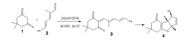
Bally and Rablen have followed up their important study of the appropriate basis sets and density functional needed to compute NMR chemical shifts1 (see this post) with this great examination of procedures for computing proton-proton coupling constants.2

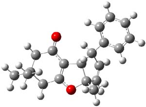
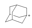
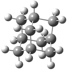
They performed a comparison of 165 experimental coupling constants from 66 small, rigid molecules with computed proton-proton coupling constants. They use a variety of basis sets and functionals. They also test whether all four components that lead to nuclear-nuclear spin coupling constants are need, or if just the Fermi contact term would suffice.

The computationally most efficient procedure, one that still provides excellent agreement with the experimental coupling constants is the following:

  1. optimize the geometry at B3LYP/6-31G(d)
  2. Calculate only the proton-proton Fermi contact terms at B3LYP/6-31G(d,p)u+1s[H]. The basis set used for computing the Fermi contact terms is unusual. The basis set for hydrogen (denoted as “u+1s[H]”) uncontracts the core functions and adds one more very compact 1s function.
  3. Scale the Fermi contact terms by 0.9155 to obtain the proton-proton coupling constants.

This methodology provides coupling constants with a mean error of 0.51 Hz, and when applied to a probe set of 61 coupling constants in 37 different molecules (including a few that require a number of conformers and thus a Boltzmann-weighted averaging of the coupling constants) the mean error is only 0.56 Hz.

Bally and Rablen supply a set of scripts to automate the computation of the coupling constants according to this prescription; these scripts are available in the supporting materials and also on the Cheshire web site. It should also be noted that the procedure described above can be performed with Gaussian-09; no other software is needed. Thus, these computations are amenable to synthetic chemists with a basic understanding of quantum chemistry.

References

(1) Jain, R.; Bally, T.; Rablen, P. R., "Calculating Accurate Proton Chemical Shifts of Organic Molecules with Density Functional Methods and Modest Basis Sets," J. Org. Chem., 2009, 74, 4017-4023, DOI: 10.1021/jo900482q.

(2) Bally, T.; Rablen, P. R., "Quantum-Chemical Simulation of 1H NMR Spectra. 2. Comparison of DFT-Based Procedures for Computing Proton-Proton Coupling Constants in Organic Molecules," J. Org. Chem., 2011, 76, 4818-4830, DOI: 10.1021/jo200513q

NMR Steven Bachrach 13 Dec 2011 6 Comments

Computed NMR spectra predicts the structure of Nobilisitine A

Nobilisitine A was isolated by Evidente and coworkers, who proposed the structure 1.1 Banwell and co-workers then synthesized the enantiomer of 1, but its NMR did not correspond to that of reported for Nobilisitine A.; the largest differences are 4.7 ppm for the 13C NMR and 0.79 ppm for the 1H NMR.2


1

Lodewyk and Tantillo3 examined seven diastereomers of 1, all of which have a cis fusion between the saturated 5 and six-member rings (rings C and D). Low energy conformations were computed for each of these diasteromers at B3LYP/6-31+G(d,p). NMR shielding constants were then computed in solvent (using a continuum approach) at mPW1PW91/6-311+G(2d,p). A Boltzmann weighting of the shielding contants was then computed, and these shifts were then scaled as described by Jain, Bally and Rablen4 (discussed in this post). The computed NMR shifts for 1 were compared with the experimental values, and the mean deviations for the 13C and 1H svalues is 1.2 and 0.13 ppm, respectively. (The largest outlier is 3.4 ppm for 13C and 0.31 for 1H shifts.) Comparison was then made between the computed shifts of the seven diasteomers and the reported spectrum of Nobilisitine A, and the lowest mean deviations (1.4 ppm for 13C and 0.21 ppm for 1H) is for structure 2. However, the agreement is not substantially better than for a couple of the other diasteomers.


2

They next employed the DP4 analysis developed by Smith and Goodman5 for just such a situation – where you have an experimental spectrum and a number of potential diastereomeric structures. (See this post for a discussion of the DP4 method.)The DP4 analysis suggests that 2 is the correct structure with a probability of 99.8%.

Banwell has now synthesized the compound with structure 2 and its NMR matches that of the original natural product.6 Thus Nobilisitine A has the structure 2.

References

(1) Evidente, A.; Abou-Donia, A. H.; Darwish, F. A.; Amer, M. E.; Kassem, F. F.; Hammoda, H. A. m.; Motta, A., "Nobilisitine A and B, two masanane-type alkaloids from Clivia nobilis," Phytochemistry, 1999, 51, 1151-1155, DOI: 10.1016/S0031-9422(98)00714-6.

(2) Schwartz, B. D.; Jones, M. T.; Banwell, M. G.; Cade, I. A., "Synthesis of the Enantiomer of the Structure Assigned to the Natural Product Nobilisitine A," Org. Lett., 2010, 12, 5210-5213, DOI: 10.1021/ol102249q

(3) Lodewyk, M. W.; Tantillo, D. J., "Prediction of the Structure of Nobilisitine A Using Computed NMR Chemical Shifts," J. Nat. Prod., 2011, 74, 1339-1343, DOI: 10.1021/np2000446

(4) Jain, R.; Bally, T.; Rablen, P. R., "Calculating Accurate Proton Chemical Shifts of Organic Molecules with Density Functional Methods and Modest Basis Sets," J. Org. Chem., 2009, DOI: 10.1021/jo900482q.

(5) Smith, S. G.; Goodman, J. M., "Assigning Stereochemistry to Single Diastereoisomers by GIAO NMR Calculation: The DP4 Probability," J. Am. Chem. Soc., 2010, 132, 12946-12959, DOI: 10.1021/ja105035r

(6) Schwartz, B. D.; White, L. V.; Banwell, M. G.; Willis, A. C., "Structure of the Lycorinine Alkaloid Nobilisitine A," J. Org. Chem., 2011, ASAP, DOI: 10.1021/jo2016899

InChIs

2: InChI=1/C17H19NO4/c19-12-3-8-1-2-18-17(8)16-10-6-15-14(21-7-22-15)5-9(10)13(20)4-11(12)16/h5-6,8,11-12,16-19H,1-4,7H2/t8-,11-,12-,16-,17-/m0/s1

InChIKey=JISHLXUXALHAET-PUYTVRRYBF

NMR Steven Bachrach 15 Nov 2011 3 Comments

cyclopenta[b]benzofuran – stereochemistry and mechanism of formation

Here is a nice example of an interesting synthesis, mechanistic explication using computation (with a bit of an unanswered question), and corroboration of the stereochemistry of the product using computed NMR shifts. Gil and Mischne1 reacted dimedone 1 with dienal 2 under Knoevenagel conditions to give, presumably, 3. But 3 is not recovered, rather the tricycle 4 is observed.

There are four stereoisomers that can be made (4a-d). Computed 13C chemical shifts at OPBE/pcS-1 (this is a basis set suggested for computing chemical shifts2) for these four isomers were then compared with the experimental values. The smallest root mean squared error is found for 4d. Better still, is that these authors utilized the DP4 method of Goodman3 (see this post), which finds that 4d agrees with the experiment with 100% probability!

Lastly, the mechanism for the conversion of 3 to 4 was examined at M06/6-31+G**. The optimized geometries of the starting material, transition state, and product are shown in Figure 1. The free energy barrier is a modest 14.5 kcal mol-1. The TS indicates a conrotatory 4πe electrocyclization. The formation of the C-O bond lags far behind in the TS. They could not identify a second transition state. It would probably be worth examining whether the product of this 4πe electrocyclization could be located, perhaps with an IRC starting from the transition state. Does this TS really connect 3 to 4?

3

TS

4

Figure 1. M06/6-31+G** optimized geometries of 3 and 4 and the transition state connecting them.

References

(1) Riveira, M. J.; Gayathri, C.; Navarro-Vazquez, A.; Tsarevsky, N. V.; Gil, R. R.; Mischne, M. P., "Unprecedented stereoselective synthesis of cyclopenta[b]benzofuran derivatives and their characterisation assisted by aligned media NMR and 13C chemical shift ab initio predictions," Org. Biomol. Chem., 2011, 9, 3170-3175, DOI: 10.1039/C1OB05109A

(2) Jensen, F., "Basis Set Convergence of Nuclear Magnetic Shielding Constants Calculated by Density Functional Methods," J. Chem. Theory Comput., 2008, 4, 719-727, DOI: 10.1021/ct800013z

(3) Smith, S. G.; Goodman, J. M., "Assigning Stereochemistry to Single Diastereoisomers by GIAO NMR Calculation: The DP4 Probability," J. Am. Chem. Soc., 2010, 132, 12946-12959, DOI: 10.1021/ja105035r

InChIs

1: InChI=1/C8H12O2/c1-8(2)4-6(9)3-7(10)5-8/h3-5H2,1-2H3
InChIKey=BADXJIPKFRBFOT-UHFFFAOYAX

2: InChI=1/C12H12O/c1-11(10-13)6-5-9-12-7-3-2-4-8-12/h2-10H,1H3/b9-5+,11-6+
InChIKey=VFBDYWDOVMUDEB-MPEOSAONBY

3: InChI=1/C20H22O2/c1-15(8-7-11-16-9-5-4-6-10-16)12-17-18(21)13-20(2,3)14-19(17)22/h4-12H,13-14H2,1-3H3/b11-7+,15-8+
InChIKey=IBEGRISKTNRVOU-YQQAFNMCBC

4d: InChI=1/C20H22O2/c1-19(2)11-15(21)17-16(12-19)22-20(3)10-9-14(18(17)20)13-7-5-4-6-8-13/h4-10,14,18H,11-12H2,1-3H3/t14-,18+,20+/m1/s1
InChIKey=VEGSTZFBNRXEAX-WNYOCNMUBZ

electrocyclization &NMR Steven Bachrach 23 Aug 2011 3 Comments

1-Adamantyl cation – Predicting its NMR spectra

What is required in order to compute very accurate NMR chemical shifts? Harding, Gauss and Schleyer take on the interesting spectrum of 1-adamantyl cation to try to discern the important factors in computing its 13C and 1H chemical shifts.1


1

To start, the chemical shifts of 1-adamtyl cation were computed at B3LYP/def2-QZVPP and
MP2/qz2p//MP2/cc-pVTZ. The root means square error (compared to experiment) for the carbon chemical shifts is large: 12.76 for B3LYP and 6.69 for MP2. The proton shifts are predicted much more accurately with an RMS error of 0.27 and 0.19 ppm, respectively.

The authors speculate that the underlying cause of the poor prediction is the geometry of the molecule. The structure of 1 was optimized at HF/cc-pVTZ, MP2/cc-pVTZ and CCSD(T)/pVTZ and then the chemical shifts were computed using MP2/tzp with each optimized geometry. The RMS error of the 12C chemical shifts are HF/cc-pVTZ: 9.55, MP2/cc-pVTZ: 5.62, and CCSD(T)/pVTZ: 5.06. Similar relationship is seen in the proton chemical shifts. Thus, a better geometry does seem to matter. The CCSD(T)/pVTZ optimized structure of 1 is shown in Figure 1.

1

Figure 1. CCSD(T)/pVTZ optimized structure of 1.

Unfortunately, the computed chemical shifts at CCSD(T)/qz2p//CCSD(T)/cc-pVTZ are still in error; the RMS is 4.78ppm for the carbon shifts and 0.26ppm for the proton shifts. Including a correction for the zero-point vibrational effects and adjusting to a temperature of 193 K to match the experiment does reduce the error; now the RMS for the carbon shifts is 3.85 ppm, with the maximum error of 6 ppm for C3. The RMS for the proton chemical shifts is 0.21ppm.

The remaining error they attribute to basis set incompleteness in the NMR computation, a low level treatment of the zero-point vibrational effects (which were computed at HF/tz2p), neglect of the solvent, and use of a reference in the experiment that was not dissolved in the same media as the adamantyl cation.

So, to answer our opening question – it appears that a very good geometry and treatment of vibrational effects is critical to accurate NMR shift computation of this intriguing molecule. Let the
computational chemist beware!

References

(1) Harding, M. E.; Gauss, J.; Schleyer, P. v. R., "Why Benchmark-Quality Computations Are Needed To Reproduce 1-Adamantyl Cation NMR Chemical Shifts Accurately," J. Phys. Chem. A, 2011, 115, 2340-2344, DOI: 10.1021/jp1103356

InChI

1: InChI=1/C10H15/c1-7-2-9-4-8(1)5-10(3-7)6-9/h7-9H,1-6H2/q+1
InChIKey=HNHINQSSKCACRU-UHFFFAOYAC

adamantane &NMR &Schleyer Steven Bachrach 18 Jul 2011 4 Comments

Structure of vannusal B

Saelli, Nicolaou, and Bagno point out in a recent article how the determination of the structure of vannusal B might have been guided by DFT computed 13C NMR chemical shifts, had they been available.1 The original structure was proposed in 1999 as 1,2 but was ultimately settled as 2 in 2010.3


1


2

The 13C NMR chemical shifts of 1 and 2 and some other alternatives were computed at M06/pcS-2//B3LYP/6-31g(d,p), where the pcS-2 basis set4 is one proposed by Jensen for computing chemical shifts. The computed chemical shifts of 1 poorly correlate with the experimental chemical shifts of vannusal B, with a low correlation coefficient of 0.9580 and a maximum error of 16.2 ppm. On the other hand, the correlation between the computed chemical shifts of 2 with the experimental values is excellent (R2=0.9948) and a maximum error of 3.0 ppm. Comparison of computed and experimental H-H coupling constants of model compounds of the “northeast” section of the molecule verified the correct structure is 2.

References

(1) Saielli, G.; Nicolaou, K. C.; Ortiz, A.; Zhang, H.; Bagno, A., "Addressing the Stereochemistry of Complex Organic Molecules by Density Functional Theory-NMR: Vannusal B in Retrospective," J. Am. Chem. Soc., 2011, 133, 6072-6077, DOI: 10.1021/ja201108a

(2) Guella, G.; Dini, F.; Pietra, F., "Metabolites with a Novel C30 Backbone from Marine Ciliates," Angew. Chem. Int. Ed., 1999, 38, 1134-1136, DOI: 10.1002/(SICI)1521-3773(19990419)38:8<1134::AID-ANIE1134>3.0.CO;2-U

(3) Nicolaou, K. C.; Ortiz, A.; Zhang, H.; Dagneau, P.; Lanver, A.; Jennings, M. P.; Arseniyadis, S.; Faraoni, R.; Lizos, D. E., "Total Synthesis and Structural Revision of Vannusals A and B: Synthesis of the Originally Assigned Structure of Vannusal B," J. Am. Chem. Soc., 2010, 132, 7138-7152, DOI: 10.1021/ja100740t

(4) Jensen, F., "Basis Set Convergence of Nuclear Magnetic Shielding Constants Calculated by Density Functional Methods," J. Chem. Theory Comput., 2008, 4, 719-727, DOI: 10.1021/ct800013z

InChI

vannusal B (2):
InChI=1/C31H46O5/c1-16(2)18-6-7-19(17(18)3)20-8-9-21-22(20)14-29(15-32)24-11-13-31(29,25(21)33)27(35)30(24)12-10-23(26(30)34)28(4,5)36/h14-15,17-21,23-27,33-36H,1,6-13H2,2-5H3/t17-,18+,19+,20+,21-,23+,24+,25+,26?,27+,29+,30-,31-/m1/s1
InChIKey=KYOBJLKAZYUEHK-GYGUSHOLBX

NMR Steven Bachrach 24 May 2011 3 Comments

Assigning a computed NMR spectrum – the case of one diastereomer

What procedure should one employ when trying to determine a chemical structure from an NMR spectrum? I have discussed a number of such examples in the past, most recently the procedure by Goodman for dealing with the situation where one has the experimental spectra of 2 diastereomers and you are trying to identify the structures of this pair.1 Now, Goodman provides an extension for the situation where you have a single experimental NMR spectrum and you are trying to determine which of a number of diasteromeric structures best accounts for this spectrum.2 Not only does this prescription provide a means for identifying the best structure, it also provides a confidence level.

The method, called DP4, works as follows. First, perform an MM conformational search of every diastereomer. Select the conformations within 10 kJ of the global minimum and compute the 13C and 1H NMR chemical shifts at B3LYP/6->31G(d,p) – note no reoptimizations! Then compute the Boltzmann weighted average chemical shift. Scale these shifts against the experimental values. You’re now ready to apply the DP4 method. Compute the error in each chemical shift. Determine the probability of this error using the Student’s t test (with mean, standard deviation, and degrees of freedom as found using their database of over 1700 13C and over 1700 1H chemical shifts). Lastly, the DP4 probability is computed as the product of these probabilities divided by the sum of the product of the probabilities over all possible diastereomers. This process is not particularly difficult and Goodman provides a Java applet to perform the DP4 computation for you!

In the paper Smith and Goodman demonstrate that in identifying structures for a broad range of natural products, the DP4 method does an outstanding job at identifying the correct diastereomer, and an even better job of not misidentifying a wrong structure to the spectrum. Performance is markedly better than the typical procedures used, like using the correlation coefficient or mean absolute error. I would strongly encourage those people utilizing computed NMR spectra for identifying chemical structures to considering employing the DP4 method – the computational method is not particularly computer-intensive and the quality of the results is truly impressive.

Afternote: David Bradley has a nice post on this paper, including some comments from Goodman.

References

(1) Smith, S. G.; Goodman, J. M., "Assigning the Stereochemistry of Pairs of Diastereoisomers Using GIAO NMR Shift Calculation," J. Org. Chem., 2009, 74, 4597-4607, DOI: 10.1021/jo900408d

(2) Smith, S. G.; Goodman, J. M., "Assigning Stereochemistry to Single Diastereoisomers by GIAO NMR Calculation: The DP4 Probability," J. Am. Chem. Soc., 2010, 132, 12946-12959, DOI: 10.1021/ja105035r

DFT &NMR Steven Bachrach 28 Sep 2010 6 Comments

Computed NMR chemical shifts with multiple standards

In order to obtain computed NMR chemical shifts, one computes the isotropic magnetic shielding tensor and subtracts this value from that computed for a reference (or standard) compound. Typically, one uses TMS as the standard. Sarotti and Pellegrinet have questioned whether this is a reasonable approach.1 Since computational methods vary in quality with methodology, basis set, geometry – one might wonder if the use of a single standard for all computed chemical shifts is the best approach.

They computed the 13C chemical shielding tensor for 50 organic compounds possessing a wide variety of functional groups and rings – a few examples are given below. They also computed the 13C chemical shielding tensor for 11 different simple organic compounds that might be used as NMR references (like TMS, benzene, methanol, and chloroform).

By comparing the computed chemical shifts obtained using the different references and then matching them with experiment, they propose a multi-reference method. For sp3 carbon atoms they propose using methanol as the reference, and for sp2 and sp carbons using benzene as the reference. With chemical shifts computed at mPW1PW91/6-311+G(2d,p)//B3LYP/6-31G(d) using the multi-reference model , the average mean difference from experiment is 2.1 ppm, less than half that found when TMS alone is used. The average RMS deviation of 4.6ppm is about half that when TMS is used as the sole standard.

Though the authors mention the solvent effect on chemical shifts, it is surprising that they did not include solvent in their calculations, especially since they are comparing to experimental chemical shifts in deuterochloroform. Nonetheless, I think this is a nice idea and further exploration of this concept (multi-reference fitting) is worth further pursuits.

References

(1) Sarotti, A. M.; Pellegrinet, S. C., "A Multi-standard Approach for GIAO 13C NMR Calculations," J. Org. Chem., 2009, 74, 7254-7260, DOI: 10.1021/jo901234h

NMR Steven Bachrach 11 May 2010 2 Comments

NMR shifts of aromatic and antiaromatic compounds using BLW

The chemical shift of the benzene proton is about 7.3ppm, significantly downfield from the range of olefinic protons (5.6-58.ppm). This is rationalized as the standard induced diatropic ring current, found in aromatic species. But what should we make of the chemical shift of the protons in cyclobutadiene at 5.8 ppm? Shouldn’t this be much further upfield?

Schleyer and Mo have applied the block localized wavefunction (BLW) technique to aromatic and antiaromatic chemical shifts.1 In BLW, self-consistent localized orbitals are produced to describe a particular resonance structure. So, for benzene, BLW describes in effect 1,3,5-cyclohexatriene, lacking any resonance energy.  When chemical shifts are computed with the BLW description, the proton chemical shift is 6.6 ppm, and is even more upfield if the geometry is optimized (in D3h symmetry) with the BLW method (δ=6.2ppm). Furthermore the NICS(0)πzz (the tensor component corresponding to the perpendicular direction evaluated in the ring center using just the π orbitals) is -36.3 for benzene and 0.0 for the D3h BLW variant, strongly indicating the role of cyclic delocalization in affecting chemical shifts.

Now for cyclobutadiene, the proton chemical shift of 5.7 ppm becomes 7.4 in the BLW case. NICS(0)πzz for cyclobutadiene is +46.9 and +1.6 in the BLW case. The problem is that typical alkenes are poor references for cyclobutadiene – when resonance is turned off, the chemical shift does move downfield – indicating the expected upfield shift for cyclobutadiene. Schleyer and Mo suggest that 3,4-dimethylenecyclobutene is a more suitable reference; its ring protons have chemical shifts of 7.65ppm.

They also describe computations of benzocyclobutadiene and tricyclobutenabenzene and offer straightforward rationalizations of their aromatic vs. antiaromatic behavior.

References

(1) Steinmann, S. N.; Jana, D. F.; Wu, J. I.-C.; Schleyer, P. v. R.; Mo, Y.; Corminboeuf, C., "Direct Assessment of Electron Delocalization Using NMR Chemical Shifts," Angew. Chem. Int. Ed., 2009, 48, 9828-9833, DOI: 10.1002/anie.200905390

InChIs

benzene: InChI=1/C6H6/c1-2-4-6-5-3-1/h1-6H
InChIKey=UHOVQNZJYSORNB-UHFFFAOYAH

cyclobutadiene: InChI=1/C4H4/c1-2-4-3-1/h1-4H
InChIKey=HWEQKSVYKBUIIK-UHFFFAOYAI

3,4-dimethylenecyclobutene: InChI=1/C6H6/c1-5-3-4-6(5)2/h3-4H,1-2H2
InChIKey=WHCRVRGGFVUMOK-UHFFFAOYAP

Aromaticity &NMR &Schleyer Steven Bachrach 04 May 2010 No Comments

A new approach to NMR structure prediction

I have written many posts on the use of computed NMR shifts as a tool for determining molecular structure, especially stereochemistry. All of these methods rely upon computing a bunch of alternative structures and then identifying the one whose chemical shifts (1H and/or 13C) match up best with experiment. Many people have been interested in the first part of this process – the “computing a bunch of alternative structures” – testing the QM method, the basis set, the selection of conformation(s), and the method for computing chemical shifts. The subject of this post is the notion of “matching up best” and comes from of a recent article by Jonathan Goodman.1

So in the typical procedure for deciding which structure (of many) best accounts for the experimental NMR spectra, the computed NMR shifts (and perhaps coupling constants) are compared to the experimental data. This comparison is done often by simply examining the correlation coefficient r between the experimental and calculated shifts. Some have used the mean absolute error between the computed and experimental shifts. Others have employed a corrected mean absolute error where scaled chemical shifts are first obtained from the plot of the calculated vs. experimental shifts, and then finding the average of the differences between these scaled shifts and the experimental ones.

Goodman suggests that oftentimes what is of interest is not really the chemical shifts of a compound but rather identifying the structure of diastereomers, and then it’s really the differences in the chemical shifts of pairs of diastereomers that are really critical in identifying which one is which. Using Goodman’s notation, suppose you have experimental NMR data on diastereomers A and B and the computed NMR shifts for structures a and b. The key is deciding does A correlate with a or b and the same for B. Goodman proposes three variants on how to compare the chemical shift differences, but I’ll show just the first, which he calls CP1. Define Δexpi as the differences in the experimental chemical shifts of the two diastereomers for nucleus i: Δexp = δAi – δBi and a similar definition for the differences in the computed shifts: Δcalc = δai – δbi. CP1 is then defined as Σ (Δexpcalc)/Σ (Δexp)2 where each sum is over the nuclei i. Goodman shows in a number of examples (some are shown below) that CP1 and its variants provides an excellent measure of when a computed structure’s chemical shifts agree with the experimental values, along with a means for noting the confidence in that assignment. These CP measures provide significantly better measures of agreement that the ones previous utilized, providing a real confidence level in assessing the quality of the prediction. I strongly urge all who are interested in the use of computed NMR in determining molecular structures to read this paper and consider adopting this approach.

References

(1) Smith, S. G.; Goodman, J. M., "Assigning the Stereochemistry of Pairs of Diastereoisomers Using GIAO NMR Shift Calculation," J. Org. Chem. 2009, 74, 4597-4607, DOI: 10.1021/jo900408d

NMR Steven Bachrach 25 Aug 2009 No Comments

Computed NMR – structure of isorunanine and hypurticin

Here are two nice examples of the use of computed spectra in identifying the structure of large molecules.

Castle and co-workers describe the synthesis of what they hoped would be runanine 1.1 However, after they had completed their synthesis, the 1H NMR spectrum of their product differed significantly from that of runanine. Further the optical rotation of 1 is -400, while that of their product is -34. Speculating on what might be the product they came up with 4 alternative structures 2-5. The 13C NMR of 1-5 were then computed by optimizing the structures at mPW1PW91/6-31G* followed by a GIAO computation at mPW1PW91/aug-cc-pVDZ with PCM (solvent is chloroform). The differences between the computed chemical shifts for 1-5 and the experimental shifts of the obtained product are summarized in Table 1. The authors conclude that their product is 5, a compound they name isorunanine.


1
(runanine)


2


3


4


5
(isorunanine)

Table 1. Average difference and maximum difference between the computed and experimental 13 C chemical shifts (ppm).

Compound

Average Δδ

Maximum Δδ

1

9.7

61.7

2

9.3

60.1

3

12.0

54.6

4

10.9

61.8

5

3.8

9.8

The authors also report the rather poor agreement between the computer spectrum of 6 and the experimental spectrum in benzene. Unfortunately, not enough details are provided to really determine where errors might be occurring. For example, there is no indication of examining multiple conformations (and those methoxy groups can rotate along with the inversion at the amine). Once again, the supporting materials, while extensive in terms of experimental NMR spectra, contain no details of the computed structures.

The structure of hypurticin 6 was determined using a comparison of computed coupling constants.2 Here the authors first assumed that four possible stereoisomers are possible 6a-d, given that the other stereocenters were determined unambiguously by experiment and biogenesis considerations. B3LYP/6-31G(d) optimization of a restricted set of conformations led to the lowest energy conformer. The coupling constants computed for these four structures indicated the closet agreement between the computed constants of 6a with experimental values. An exhaustive search of the conformational space of each of these diastereomers at B3LYP/DGDZVP followed by Boltzmann weighting of the coupling constants confirmed that 6a is the structure of hypurticin.

References

(1) Nielsen, D. K.; Nielsen, L. L.; Jones, S. B.; Toll, L.; Asplund, M. C.; Castle, S. L., "Synthesis of Isohasubanan Alkaloids via Enantioselective Ketone Allylation and Discovery of an Unexpected Rearrangement," J. Org. Chem. 2009, 74, 1187-1199, DOI: 10.1021/jo802370v.


(2) Mendoza-Espinoza, J. A.; Lopez-Vallejo, F.; Fragoso-Serrano, M.; Pereda-Miranda, R.; Cerda-Garcia-Rojas, C. M., "Structural Reassignment, Absolute Configuration, and Conformation of Hypurticin, a Highly Flexible Polyacyloxy-6-heptenyl-5,6-dihydro-2H-pyran-2-one," J. Nat. Prod. 2009, 72, 700-708, DOI: 10.1021/np800447k.

NMR Steven Bachrach 29 Jun 2009 No Comments

« Previous PageNext Page »