Archive for the 'NMR' Category

Computing 1H NMR chemical shifts

Computed NMR spectra have been a major theme of the blog (see these posts). General consensus is that they can be enormously helpful in characterizing structures and stereochemistry, but there has been a nagging sense that one needs to use very large basis sets to get reasonable accuracies.

Bally and Rablen1 now confront that claim and suggest instead that quite modest basis sets along with a number of flavors of DFT can provide very good 1H NMR shifts. They examined 80 organic molecules spanning a variety of functional groups. A key feature is that these molecules exist as a single conformation or their conformational distribution is dominated by one conformer. This avoids the need of computing a large number of conformers and taking a Boltzman average of their shifts – a task that would likely require a much larger basis set than what they hope to get away with.

The most important conclusion: the WP04 functional,2 developed by Cramer to predict proton spectra, with the very small 6-31G(d,p) basis set and incorporation of the solvent through PCM provides excellent cost/benefit performance. The rms error of the proton chemical shifts is 0.198 ppm, and this can be reduced to 0.140 ppm with scaling. The 6-31G(d) basis set is even better if one uses a linear scaling; its error is only 0.120 ppm. B3LYP/6-31G(d,p) has an rms only somewhat worse. Use of aug-cc-pVTZ basis sets, while substantially more time consuming, provides inferior predictions.

The authors contend that this sort of simple DFT computation, affordable for many organic systems on standard desktop PCs, should be routinely done, especially in preference to increment schemes that are components of some drawing programs. And if a synthesis group does not have the tools to do this sort of work, I’m sure there are many computational chemists that would be happy to collaborate!

References

(1) Jain, R.; Bally, T.; Rablen, P. R., "Calculating Accurate Proton Chemical Shifts of Organic Molecules with Density Functional Methods and Modest Basis Sets," J. Org. Chem. 2009, DOI: 10.1021/jo900482q.

(2) Wiitala, K. W.; Hoye, T. R.; Cramer, C. J., "Hybrid Density Functional Methods Empirically Optimized for the Computation of 13C and 1H Chemical Shifts in Chloroform Solution," J. Chem. Theory Comput. 2006, 2, 1085-1092, DOI: 10.1021/ct6001016

DFT &NMR Steven Bachrach 15 Jun 2009 3 Comments

Hexacylinol (again)

One more nail in the coffin of the widely disputed Le Clair structure of hexacyclinol is provided by the B97-2/cc-pVTZ/B3LYP/6-31G(d,p) computed proton and 13C NMR for the two “structures” (see my previous blog post for structures and background). These computations1 are at a more rigorous level than those performed by Rychnovsky, and the addition of the proton spectrum helps clearly settle this issue. Rychnovsky’s structure is the correct one – the mean absolute error between the experimental and computed structure is half that for Rychnovsky structure. The computed coupling constants also are in much better agreement with the Rychnovsky structure. So, Bagno’s contribution accomplishes, I hope, two things: (1) convinces everyone that DFT NMR spectra can be an important tool in identifying natural product structure and (2) closes the book on hexacylinol!

References

(1) Saielli, G.; Bagno, A., "Can Two Molecules Have the Same NMR Spectrum? Hexacyclinol Revisited," Org. Lett. 2009, 11, 1409-1412, DOI: 10.1021/ol900164a.

DFT &hexacyclinol &NMR Steven Bachrach 18 Mar 2009 1 Comment

Computed NMR spectra to identify the structure of Samoquasine A

Here’s another nice example of computed NMR spectra being
used to identify complex organic structures.1

An alkaloid isolated from the custard apple tree was assigned the structure 1 and christened with the name samoquasine A.2 Two years later, the authors determined that samoquasine A was actually identical to perlolidine 2.3 Independent synthesis of the compound with structure 1 showed that its properties were not identical to that of samoquasine A.4,5 The properties of perlolidine were then found to differ from that of samoquasine A,4 leaving a void as to just what is the structure of samoquasine A.

Given that compounds 1 and the related compounds 3 and 4 had been prepared and their NMR spectra obtained, Timmons and Wipf1 decided to compute the 13C NMR spectra of 48 related compounds at B3LYP/6-311+G(2d,p)//B3LYP/6-31G(d). The mean absolute difference between the computed and experimental chemical shifts for 1, 3 and 4 are less than 2 ppm. Of the remaining 45 compounds, the one whose chemical shifts match best with that of samoquasine A is 2, with a mean absolute deviation of 1.8 ppm. This agreement supports the contention that samoquasine A and perlolidine are in fact identical. The authors contend that the experimental data used to conjecture that they were not identical is in fact faulty.

References

(1) Timmons, C.; Wipf, P., "Density Functional Theory Calculation of 13C NMR Shifts of Diazaphenanthrene Alkaloids: Reinvestigation of the Structure of Samoquasine A," J. Org. Chem., 2008, 73, 9168-9170, DOI: 10.1021/jo801735e.

(2) Morita, H.; Sato, Y.; Chan, K.-L.; Choo, C.-Y.; Itokawa, H.; Takeya, K.; Kobayashi, J. i., "Samoquasine A, a Benzoquinazoline Alkaloid from the Seeds of Annona squamosa," J. Nat. Prod., 2000, 63, 1707-1708, DOI: 10.1021/np000342i.

(3) Morita, H.; Sato, Y.; Chan, K.-L.; Choo, C.-Y.; Itokawa, H.; Takeya, K.; Kobayashi, J. i., "Samoquasine A, a Benzoquinazoline Alkaloid from the Seeds of Annona squamosa," J. Nat. Prod., 2002, 65, 1748-1748, DOI: 10.1021/np0204343.

(4) Yang, Y.-L.; Chang, F.-R.; Wu, Y.-C., "Total synthesis of 3,4-dihydrobenzo[h]quinazolin-4-one
and structure elucidation of perlolidine and samoquasine A," Tetrahedron Letters, 2003, 44, 319-322, DOI: 10.1016/S0040-4039(02)02577-7.

(5) Chakrabarty, M.; Sarkara, S.; Harigaya, Y., "An Expedient Synthesis of Benzo[h]quinazolin-4(3H)-one: Structure of Samoquasine A Revisited," Synthesis, 2003, 2292-2294, DOI: 10.1055/s-2003-42409.

InChIs

1: InChI=1/C12H8N2O/c15-12-10-6-5-8-3-1-2-4-9(8)11(10)13-7-14-12/h1-7H,(H,13,14,15)/f/h14H
InChIKey=BJVYARVTSUNBMW-YHMJCDSICO

2: InChI=1/C12H8N2O/c15-12-10-7-14-11-4-2-1-3-9(11)8(10)5-6-13-12/h1-7H,(H,13,15)/f/h13H
InChIKey=ULIAUQBOGQCMQM-NDKGDYFDCS

3: InChI=1/C12H8N2O/c15-12-10-6-5-8-3-1-2-4-9(8)11(10)7-13-14-12/h1-7H,(H,14,15)/f/h14H
InChIKey=JFJSVLSRVOIBPG-YHMJCDSICJ

4: InChI=1/C12H8N2O/c15-12-11-9(7-13-14-12)6-5-8-3-1-2-4-10(8)11/h1-7H,(H,14,15)/f/h14H
InChIKey=BGYAATPHYRJYHZ-YHMJCDSICO

DFT &NMR Steven Bachrach 15 Jan 2009 No Comments

Predicting the 13C NMR of elatenyne

At the Spring ACS meeting in New Orleans this past April, Jonathan Goodman told me about his group’s new work on computed NMR spectra. This study has now appeared.1 The new aspect of this work is taking on compounds with significant conformational flexibility. They first examined the bifuranyl- and pyranopyrans acetals 1-6.


1


2


3


4


5


6

They identified all energetically low-lying conformers through a Monte Carlo MM search, and reoptimized the structures at B3LYP/6-31G**. PCM energies, with the dielectric set to 4.81 to simulate CHCl3, were obtained using the gas-phase geometries. 13C NMR shifts were then computed and weighted based on Boltzmann averaging. Compounds 4-7 require consideration of 4, 5, or 7 conformations, respectively, to account for at least 95% of the population. (The lowest energy conformation for each compound is displayed in Figure 1.) The computed 13C NMR chemical shifts were then compared against the experimental values that have been obtained for 5 of these 6 compounds, though it was not known which experimental spectra corresponds with which structure. Based on the correlation coefficients and the mean and maximum values of the differences between the calculated and experimental shifts allows for identification of the structure that corresponds to each of the five spectra.

1

2

3

4

5

6

Figure 1. B3LYP/6-31G** minimum energy conformation of 1-6.1

They next took on the absolute structure of elatenyne 7. This compound has been isolated and its 13C NMR obtained nd interpreted.2 The compound can exist in one of 32 different diastereomers. Instead of having to stereospecifically synthesize each diastereomer, obtain its NMR spectra and compare with the natural product, Goodman suggests that one can compute the 13C NMR spectra for each isomer, identified the likely candidates, and synthesize just those if verification is necessary. So, they computed the spectra of all 32 isomers following the above prescription. The chemical shifts for the carbons bearing bromine have relatively large errors, though these can by systematically corrected by reducing the computed values by about 20 ppm. Comparison of the computed spectra of each isomer with the experimental spectra gave three possible isomers (7a-c, see Figure 2) with very strong correlation coefficients. Examination of the men value of the difference of the computed chemical shifts with the experimental values (not including the carbon atoms with a bromine attached) suggests one more possibility 7d (see Figure 2).


7

7a

7b

7c

7d

Figure 2. B3LYP/6-31G** minimum energy conformation of 7a-d.1

References

(1) Smith, S. G.; Paton, R. S.; Burton, J. W.; Goodman, J. M., "Stereostructure Assignment
of Flexible Five-Membered Rings by GIAO 13C NMR Calculations: Prediction of the Stereochemistry of Elatenyne," J. Org. Chem., 2008, DOI: 10.1021/jo8003138.

(2) Sheldrake, H. M.; Jamieson, C.; Burton, J. W., “The Changing Faces of Halogenated Marine Natural Products: Total Synthesis of the Reported Structures of Elatenyne and an Enyne from Laurencia majuscula,” Angew. Chem. Int. Ed., 2006, 45, 7199-7202, DOI: 10.1002/anie.200602211

InChIs

1-3: InChI=1/C10H18O4/c1-11-9-5-3-8-7(13-9)4-6-10(12-2)14-8/h7-10H,3-6H2,1-2H3

4-6: InChI=1/C10H18O4/c1-11-9-5-3-7(13-9)8-4-6-10(12-2)14-8/h7-10H,3-6H2,1-2H3

7: InChI=1/C15H20Br2O2/c1-3-5-6-7-13-11(17)9-15(19-13)14-8-10(16)12(4-2)18-14/h1,5-6,10-15H,4,7-9H2,2H3/b6-5-
InChIKey=SKSTYQSRJPCZSW-WAYWQWQTBQ

DFT &NMR Steven Bachrach 27 May 2008 1 Comment

Assigning the structure of obtusallenes using computed NMR

Here’s another interesting application of computed NMR spectra to resolve the structure of natural products. Braddock and Rzepa have examined obtusallenes V (1), VI (2) and VII (3).1 The geometries were optimized at mPW1PW91/6-31G(d,p) and the chemical shifts were obtained at this level and using the aug-cc-pVDZ basis set. The larger basis reduces the error and no statistical correction need be applied. The coordinates of these compounds are available through this web-enhanced object of the paper.


1


2


3

The confusion in these structures relates to the position of the halide attachments. For 1 and 2, the problem is which halide (Br or Cl) is at C-7 and C-13. The original structures proposed had these halogens switched from what I’ve drawn, and the correlation between the computed chemical shifts for these original structures and the experiment shows significant deviation: a mean deviation of 1.42 ppm for 1 and 1.67 ppm for 2. Using the structures shown above, along with switching the assigned 13C chemical shifts gives much better agreement between the computed and experimental values; the mean deviation is 1.15 ppm for both 1 and 2. Unfortunately the stereochemistry about the allene cannot be determined using NMR – the two different isomers have similar chemical shifts. Similarly, the structure of 3 is predicted as shown above, though the experiment reported only some of the chemical shifts so some uncertainty remains.

References

(1) Braddock, D. C.; Rzepa, H. S., "Structural Reassignment of Obtusallenes V, VI, and VII by GIAO-Based Density Functional Prediction," J. Nat. Prod., 2008, DOI: 10.1021/np0705918.

InChIs

1: InChI=1/C15H18Br3ClO3/c1-8-14(18)12-6-13(17)15(22-12)7-10(19)11(21-15)5-9(20-8)3-2-4-16/h3-4,8-14H,5-7H2,1H3/t2-,8-,9+,10+,11-,12+,13-,14-,15-/m0/s1
InChIKey = PVIUYMGCQVXTIT-JUHTWQEGBT

2: InChI=1/C15H19Br2ClO3/c1-9-14(17)12-4-5-15(20-12)8-11(18)13(21-15)7-10(19-9)3-2-6-16/h3,6,9-14H,4-5,7-8H2,1H3/t2-,9-,10+,11+,12+,13-,14-,15-/m0/s1
InChIKey = WPEZFVRVOYPLJW-LXJGPXSEBA

3: InChI=1/C15H20Br3ClO3/c1-8-15(18)14-6-10(17)13(22-14)7-11(19)12(20)5-9(21-8)3-2-4-16/h3-4,8-15,20H,5-7H2,1H3/t2-,8-,9+,10-,11+,12-,13-,14+,15-/m0/s1
InChIKey = QTZNVLUNNGQAFG-SOAHCKLOBC

NMR Steven Bachrach 24 Mar 2008 1 Comment

NICS Scan

Professor Amnon Stanger sent me an email with a couple of comments concerning points made in my book. I take up the first of his comments here. Stanger has pointed me to what looks to be the true first study evaluating NICS on a grid of points, done by Klod and Kleinpeter in 2001.1 They computed NICS values on a 3-D grid and then created iso-chemical-shielding surfaces to pictorially represent the shielding and deshielding zones (or cones) created by π-bonds (like ethane and ethyne) and aromatic compounds. In a similar vein, Lazzeretti2 evaluated the out-of-plane component of the magnetic shielding tensor (σ||) on a 2-D grid to demonstrate the shielding and deshielding zones of π-systems, particularly aromatic systems. These plots clearly demonstrate the “cones” one typically finds in introductory organic texts to explain NMR effects.

(1) Klod, S.; Kleinpeter, E., “Ab Initio Calculation of the Anisotropy Effect of Multiple Bonds and the Ring Current Effect of Arenes—Application in Conformational and Configurational Analysis,” Chem. Soc., Perkin Trans. 2, 2001, 1893-1898, DOI: 0.1039/b009809o.

(1) Viglione, R. G.; Zanasi, R.; Lazzeretti, P., “Are Ring Currents Still Useful to Rationalize the Benzene Proton Magnetic Shielding?,” Org. Lett., 2004, 6, 2265-2267, DOI: 10.1021/ol049200w.

NMR Steven Bachrach 27 Nov 2007 No Comments

Predicting NMR chemical shifts of penam β-lactams

Cramer and Hoye have applied DFT computations to the predictions of both protons and carbon NMR chemical shifts in penam β-lactams1 using the procedure previously described in my blog post Predicting NMR chemical shifts. They examined the compounds 1-8 by optimizing low energy conformers at B3LYP/6-31G(d) with IEFPCM (solvent=chloroform). The chemical shifts were then computed using these geometries with the larger 6-311+G(2d,p) basis set and four different functionals: B3LYP, PBE1 and the two specific functionals designed to produce proton and carbon chemical shifts: WP04 and WC04.

A number of interesting results are reported. First, all three functionals do a fine job in predicting the proton chemical shifts of 1-8, with WP04 slightly better than the other two.On the other hand, all three methods fail to predict the carbon chemical shifts of 1-3, though B3LYP and PBE1 do correctly identify 5-8. The failure of WC04 is surprising, especially since dimethyl disulfide was used in the training set. They also noted that WP04 using just the minimum energy conformation (as opposed to a Boltzmann averaged chemical shift sampled from many low energy conformers) did correctly identify lactams 1-4. This is helped by the fact that the lowest energy conformer constituted anywhere form 37% to 68% of the energy-weighted population.

References


(1) Wiitala, K. W.; Cramer, C. J.; Hoye, T. R., “Comparison of various density functional methods for distinguishing stereoisomers based on computed 1H or 13C NMR chemical shifts using diastereomeric penam ?-lactams as a test set,” Mag. Reson. Chem., 2007, 45, 819-829, DOI: 10.1002/mrc.2045.

InChIs

1: InChI=1/C18H17NO5S/c1-18(2)14(17(23)24-3)19-15(22)11(16(19)25-18)10-12(20)8-6-4-5-7-9(8)13(10)21/h4-7,10-11,14,16H,1-3H3/t11-,14+,16+/m0/s1

5: InChI=1/C17H15NO5S/c1-17(2)13(16(22)23)18-14(21)10(15(18)24-17)9-11(19)7-5-3-4-6-8(7)12(9)20/h3-6,9-10,13,15H,1-2H3,(H,22,23)/t10-,13+,15+/m0/s1

Cramer &DFT &NMR Steven Bachrach 22 Oct 2007 No Comments

Predicting the structure of artarborol

Here’s one more nice application of computationally-derived NMR chemical shifts towards solving a structure. Fattarusso and co-workers1 identified a component of wormwood called artarborol. COSY and ROESY experiments allowed for deducing four possible diasereomeric structures of artarborol, 1-4.

They then took two computational approaches towards resolving the structure. First, they performed an MM search for low energy conformers of 1-4. These conformers were then screened for those having a dihedral angle of around 90° for the C-8 and C-9 protons, due to a low couple constant for between these protons. Only conformers of 1 and 3 satisfied this criterion. An intense couple of the H-1 and H-5 protons indicated a transannular arrangement, and only conformers of 1 satisfy this criterion.

The second computational approach was to optimize some of the low energy conformers of 1 and 3 at mPW1PW91/6-31G(d,p) and compute their 13C chemical shifts. The five low energy conformers, two of 1 and three of 3, are shown in Figure 1. The resulting chemical shifts were averaged according to a Boltzmann distribution. These computed chemical shifts were then fit against the experimental values. The correlation factor for the computed shifts for 1 (r2=0.9997) was much better than that of 3 (r2=0.9713). The average deviation of the chemical shifts (after being corrected using the fitting procedure from the above correlation) was only 0.8ppm for 1 but 2ppm for 3. They therefore conclude that the structure of artarborol is 1.

1a
xyz

1b
xyz


3a
xyz

3b
xyz

3c
xyz

Figure 1. mPW1PW91 optimized conformations of possible artarborol diasteromers.1

References

(1) Fattorusso, C.; Stendardo, E.; Appendino, G.; Fattorusso, E.; Luciano, P.; Romano, A.; Taglialatela-Scafati, O., "Artarborol, a nor-Caryophyllane Sesquiterpene Alcohol from Artemisia arborescens. Stereostructure Assignment through Concurrence of NMR Data and Computational Analysis," Org. Lett., 2007, 9, 2377-2380, DOI: 10.1021/ol070803s.

(2) I thank Professor Ernesto Fattorusso for supplying me with the optimized coordinates of these compounds.

InChI

1: InChI=1/C14H24O2/c1-13(2)8-9-10(13)6-7-14(3)12(16-14)5-4-11(9)15/h9-12,15H,4-8H2,1-3H3/t9-,10-,11-,12-,14+/m0/s1

DFT &NMR Steven Bachrach 13 Aug 2007 No Comments

Predicting NMR chemical shifts

Another three applications of computed NMR chemical shifts towards structure identification have appeared, dealing with carbohydrates and natural products.

Prediction of NMR Signals of Carbohydrates

The study by Cramer and Hoye1 investigates identification of diastereomers with NMR, in particular, identification of cis and trans isomers of 2-methyl- (1), 3-methyl- (2), and 4-methylcyclohexanol (3). The study discusses the ability of different DFT methods to predict the chemical shifts of these alcohols in regard to distinguishing their different configurations. An interesting twist is that they have developed a functional specifically suited to predict proton chemical shifts and a second functional specifically for predicting carbon chemical shifts.2

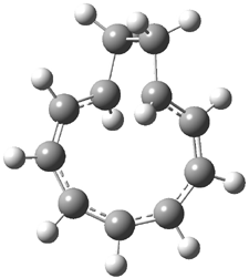
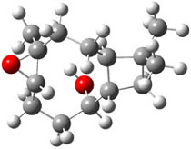
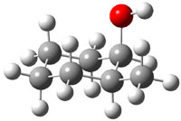
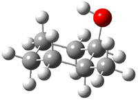
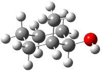
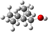
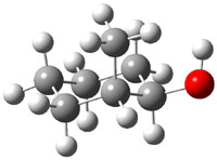
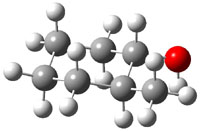
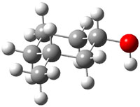
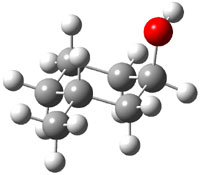
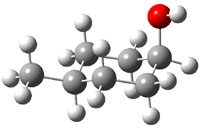
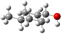
The approach they take was first to optimize the six different conformations for each diastereomer including solvent (chloroform). They chose to optimize the structures at B3LYP/6-311+G(2d,p) with PCM. The six conformers (notice the axial/equatorial relationships, along with the position of the alcohol hydrogen) of 1c are presented in Figure 1. Chemical shifts were then obtained with a number of different methods, weighting them according to a Boltzmann distribution.

0.0
xyz

0.20
xyz

0.73
xyz

1.23
xyz

1.56
xyz

1.85
xyz

Figure 1. PCM/B3LYP/6-311+G(2d,p) optimized structures of the conformers of 1c. Relative energies (kcal mol-1) are listed for each isomer.

Now a brief digression into how they developed their modified functional.2 They define the exchange-correlation functional (see Chapter 1.3.1 of my book – or many other computational chemistry books!) as

      Exc = P2Ex(HF) + P3ΔEx(B) + P4Ex(LSDA) + P5ΔEc(LYP) + P6Ec(LSDA)

where the Ps are parameters to be fit and Ex(HF) is the Hartree-Fock exchange energy, ΔEx(B) is the Becke gradient correction to the local spin-density approximation (LSDA), Ex(LSDA) is the exchange energy, ΔEc(LYP) is the Lee-Yang-Parr correction to the LSDA correlation energy, and Ec(LSDA) is the LSDA correlation energy. Chemical shifts were computed for proton and carbon, and the parameters P were adjusted (between 0 and 1) to minimize the error in the predicted chemical shifts from the experimental values. A total of 43 different molecules were used for this fitting procedure. The values of the parameters are given for the carbon functional (WC04), the proton functional (WP04) and B3LYP (as a reference) in Table 1. Note that there is substantial difference in the values of the parameter among these three different functionals.

Table 1. Values of the parameters P for the functionals WC04, WP04, and B3LYP.


P2

P3

P4

P5

P6


WC04

0.7400

0.9999

0.0001

0.0001

0.9999

WP04

0.1189

0.9614

0.999

0.0001

0.9999

B3LYP

0.20

0.72

0.80

0.81

1.00


Now, the computed proton and carbon chemical shifts using 4 different functions (B3LYP, PBE1, MP04, and WC04) for 1-3 were compared with the experiment values. This comparison was made in a number of different ways, but perhaps most compellingly by looking at the correlation coefficient of the computed shifts compared with the experimental shifts. This was done for each diastereomer, i.e. the computed shifts for 2c and 2t were compared with the experimental shifts of both 2c and 2t. If the functional works well, the correlation between the computed and experimental chemical shifts of 2c (and 2t) should be near unity, while the correlation between the computed shifts of 2c and the experimental shifts of 2t should be dramatically smaller than one. This is in fact the case for all three functionals. The results are shown in Table 2 for B3LYP and WP04, with the later performing slightly better. The results for the carbon shifts are less satisfactory; the correlation coefficients are roughly the same for all comparisons with B3LYP and PBE1, and WC04 is only slightly improved.
Nonetheless, the study clearly demonstrates the ability of DFT-computed proton chemical shifts to discriminate between diasteromers.

Table 2. Correlation coefficients between the computed and experimental proton chemical shifts.a


2ccomp
(1.06)
xyz

2tcomp
(0.0)
xyz


2cexp
 

2texp

0.9971
0.9985

0.8167
0.8098

0.8334
0.9050

0.9957
0.9843


3c
(0.0)
xyz

3t
(0.63)
xyz


3cexp
 

3texp

0.9950
0.9899

0.8856
0.9310

0.8763
0.8717

0.9990
0.9979


4c
(0.54)
xyz

4t
(0.0)
xyz


4cexp
 

4texp

0.9993
0.9975

0.8744
0.8675

0.8335
0.9279

0.9983
0.9938


aPCM/B3LYP/6-311+G(2d,p)//PCM/ B3LYP/6-31G(d) in regular type and PCM/WP04/6-311+G(2d,p)//PCM/ B3LYP/6-31G(d) in italic type. Relative energy (kcal mol-1) of the most favorable conformer of each diastereomer is given in parenthesis.

Predicting NMR of Natural Products

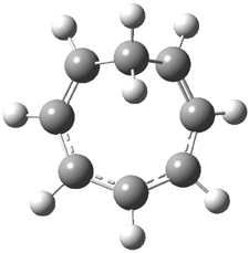
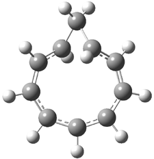
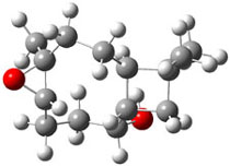
Bagno has a long-standing interest in ab initio prediction of NMR. In a recent article, his group takes on the prediction of a number of complex natural products.3 As a benchmark, they first calculated the NMR spectra of strychnine (4) and compare it with its experimental spectrum. The optimized PBE1PBE/6-31G(d,p) geometry of 4 is drawn in Figure 2. The correlation between the computed NMR chemical shifts for both 1H and 13C is quite good, as seen in Table 3. The corrected mean average errors are all very small, but Bagno does point out that four pairs of proton chemical shifts and three pairs of carbon chemical shifts are misordered.

Strychnine
4

Figure 2. PBE1PBE/6-31G(d,p) geometry of strychnine 4.3

Table 3. Correlation coefficient and corrected mean average error
(CMAE) between the computed and experiment chemical shifts of 4.


δ(1H)

δ(13C)

method

r2

CMAE

r2

CMAE

B3LYP/cc-pVTZ

0.9977

0.07

0.9979

1.4

PBE1PBE/cc-pVTZ

0.9974

0.08

0.9985

0.9


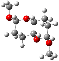
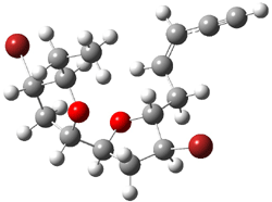
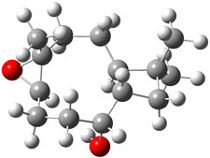
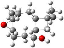
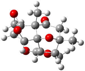
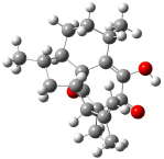
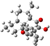
The study of the sesquiterpene carianlactone (5) demonstrates the importance of including solvent in the NMR computation. The optimized B3LYP/6-31G(d,p) geometry of 5 is shown in Figure 3, and the results of the comparison of the computed and experimental chemical are listed in Table 4. The correlation coefficient is unacceptable when the x-ray structure is used. The agreement improves when the gas phase optimized geometry is employed, but the coefficient is still too far from unity. However, optimization using PCM (with the solvent as pyridine to match experiments) and then computing the NMR chemical shifts in this reaction field provides quite acceptable agreement between the computed and experimental chemical shifts.

Corianlactone 5

Figure 3. B3LYP/6-31G(d,p) geometry of carianlactone 5.3

Table 4. Correlation coefficient and corrected mean average error (CMAE) between
the computed and experiment chemical shifts of 5.


δ(1H)

δ(13C)

geometry

r2

CMAE

r2

CMAE

X-ray

0.9268

0.23

0.9942

3.1

B3LYP/6-31G(d,p)

0.9513

0.19

0.9985

1.6

B3LYP/6-31G(d,p) + PCM

0.9805

0.11

0.9990

1.2


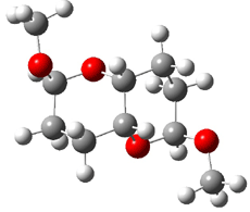
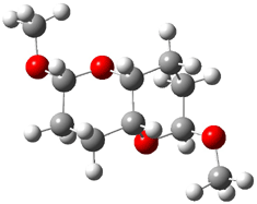
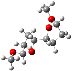
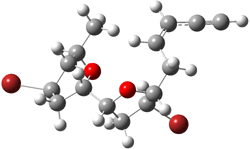
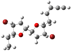
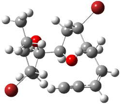
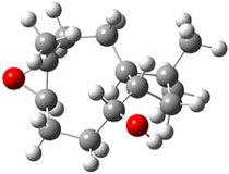
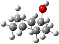
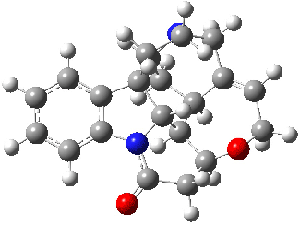
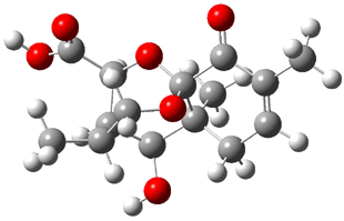
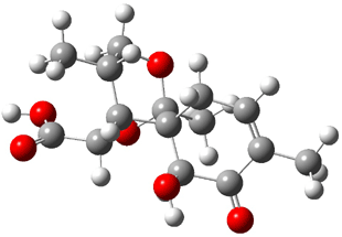
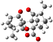
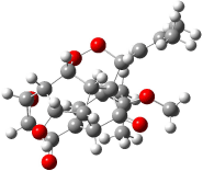
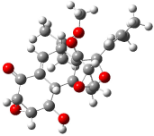
Lastly, Bagno took on the challenging structure of the natural product first identified as boletunone B (6a).4 Shortly thereafter, Steglich reinterpreted the spectrum and gave the compound the name isocyclocalopin A (6b).5 A key component of the revised structure was based on the δ 0.97 ppm signal that they assigned to a methyl above the enone group, noting that no methyl in 6a should have such a high field shift.

Bagno optimized the structures of 6a and 6b at B3LYP/6-31G(d,p), shown in Figure 4. The NMR spectra for 6a and 6b were computed with PCM (modeling DMSO as the solvent). The correlation coefficients and CMAE are much better for the 6b model than for the 6a model., supporting the reassigned structure. However, the computed chemical shift for the protons of the key methyl group in question are nearly identical in the two proposed structures: 1.08 ppm in 6a and 1.02 ppm in 6b. Nonetheless, the computed chemical shifts and coupling constants of 6b are a better fit with the experiment than those of 6a.

boletunone B 6a

isocyclocalopin A 6b

Figure 4. B3LYP/6-31G(d,p) geometry of the proposed structures of Boletunone B, 6a and 6b.3

Table 5. Correlation coefficient and corrected mean average error (CMAE) between the computed (B3LYP/6-31G(d,p) + PCM) and experiment chemical shifts of 6a and 6b.


δ(1H)

δ(13C)

structure

r2

CMAE

r2

CMAE

6a

0.9675

0.22

0.9952

3.7

6b

0.9844

0.15

0.9984

1.9


In a similar vein, Nicolaou and Frederick has examined the somewhat controversial structure of maitotxin.6 For the sake of brevity, I will not draw out the structure of maitotxin; the interested reader should check out its entry in wikipedia. The structure of maitotoxin has been extensively studied, but in 2006, Gallimore and Spencer7 questioned the stereochemistry of the J/K ring juncture. A fragment of maitotoxin that has the previously proposed stetreochemistry is 7. Gallimore and Spencer argued for a reversed stereochemistry at this juncture (8), one that would be more consistent with the biochemical synthesis of the maitotoxin. Nicolaou noted that reversing this stereochemistry would lead to other stereochemical changes in order for the structure to be consistent with the NMR spectrum. Their alternative is given as 9.

7

8

9

Nicolaou and Freferick computed 13C NMR of the three proposed fragments 7-9 at B3LYP/6-31G*; unfortunately they do not provide the coordinates. They benchmark this method against brevetoxin B, where the average error is 1.24 ppm, but they provide no error analysis – particularly no regression so that corrected chemical shift data might be employed. The best agreement between the computed and experimental chemical shifts is for 7, with average difference of 2.01 ppm. The differences are 2.85 ppm for 8 and 2.42 ppm for 9. These computations support the original structure of maitotoxin. The Curious Wavefunction blog discusses this topic, with an emphasis on the possible biochemical implication.

References

(1) Wiitala, K. W.; Al-Rashid, Z. F.; Dvornikovs, V.; Hoye, T. R.; Cramer, C. J., "Evaluation of Various DFT Protocols for Computing 1H and 13C Chemical Shifts to Distinguish Stereoisomers: Diastereomeric 2-, 3-, and 4-Methylcyclohexanols as a Test Set," J. Phys. Org. Chem. 2007, 20, 345-354, DOI: 10.1002/poc.1151

(2) Wiitala, K. W.; Hoye, T. R.; Cramer, C. J., "Hybrid Density Functional Methods Empirically Optimized for the Computation of 13C and 1H Chemical Shifts in Chloroform Solution," J. Chem. Theory Comput. 2006, 2, 1085-1092, DOI: 10.1021/ct6001016

(3) Bagno, A.; Rastrelli, F.; Saielli, G., "Toward the Complete Prediction of the 1H and 13C NMR Spectra of Complex Organic Molecules by DFT Methods: Application to Natural Substances," Chem. Eur. J. 2006, 12, 5514-5525, DOI: 10.1002/chem.200501583

(4) Kim, W. G.; Kim, J. W.; Ryoo, I. J.; Kim, J. P.; Kim, Y. H.; Yoo, I. D., "Boletunones A and B, Highly Functionalized Novel Sesquiterpenes from Boletus calopus," Org. Lett. 2004, 6, 823-826, DOI: 10.1021/ol049953i

(5) Steglich, W.; Hellwig, V., "Revision of the Structures Assigned to the Fungal Metabolites Boletunones A and B," Org. Lett. 2004, 6, 3175-3177, DOI: 10.1021/ol048724t.

(6) Nicolaou, K. C.; Frederick, M. O., "On the Structure of Maitotoxin," Angew. Chem. Int. Ed., 2007, 46, 5278-5282, DOI: 10.1002/anie.200604656.

(7) Gallimore, A. R.; Spencer, J. B., "Stereochemical Uniformity in Marine Polyether Ladders – Implications for the Biosynthesis and Structure of Maitotoxin," Angew. Chem. Int. Ed. 2006, 45, 4406-4413, DOI: 10.1002/anie.200504284.

InChI

1: InChI=1/C7H14O/c1-6-4-2-3-5-7(6)8/h6-8H,2-5H2,1H3
2: InChI=1/C7H14O/c1-6-3-2-4-7(8)5-6/h6-8H,2-5H2,1H3
3: InChI=1/C7H14O/c1-6-2-4-7(8)5-3-6/h6-8H,2-5H2,1H3
4: InChI=1/C21H22N2O2/c24-18-10-16-19-13-9-17-21(6-7-22(17)11-12(13)5-8-25-16)14-3-1-2-4-15(14)23(18)20(19)21/h1-5,13,16-17,19-20H,6-11H
5: InChI=1/C14H14O6/c1-12-2-6(15)8-13(4-18-13)9-10(19-9)14(8,20-12)7-5(12)3-17-11(7)16/h5,7-10H,2-4H2,1H3/t5-,7-,8?,9+,10+,12+,13?,14-/m1/s1
6a: InChI=1/C15H20O6/c1-7-4-5-14(3)12(17)9-8(2)6-20-15(14,11(7)16)21-10(9)13(18)19/h4,8-10,12,17H,5-6H2,1-3H3,(H,18,19)/t8-,9+,10+,12+,14-,15-/m1/s1
6b: InChI=1/C15H20O6/c1-7-4-5-15(12(17)10(7)16)9-8(2)6-20-14(15,3)21-11(9)13(18)19/h4,8-9,11-12,17H,5-6H2,1-3H3,(H,18,19)/t8?,9-,11?,12+,14-,15-/m1/s1

Cramer &DFT &NMR Steven Bachrach 01 Aug 2007 1 Comment

Predicting the Structure of Hexacyclinol

In Chapter 1.6.2 we discuss computed NMR spectra, and in particular note some successes in correlating predicted chemical shifts with experiment values. Recently, Rychnovsky took the next logical step, utilizing computational methods to predict the NMR spectrum of a compound whose structure was in doubt.

Hexacyclinol was isolated from Panus Rudis, a type of mushroom. Based on spectroscopic studies, Gräfe proposed 1 as its structure.1 Le Clair claimed to have synthesized a substance with this structure in 2006.2 This article became a cause célèbre in the blogosphere,3 with serious doubts cast upon the veracity of the author and his claims.

Rychnovsky4 doubted that the molecule actually possessed the unusual structure of 1. Since the actual structure was unknown, he proposed to compute the NMR shifts based on the optimized structure of 1 and compare them with the experimental values. Given the very large size of hexacyclinol, the computational approach would have to be rather limited. Therefore, whatever (small) method was to be employed would have to be tested for adequate predictive performance with known compounds. Rychnovsky selected the three diterpenes elisapterosin B 2, elisabethin A 3, and maoecrystal V 4 to benchmark his computations. His computational approach was to first utilize a Monte Carlo search with the MMFF force field to identify low lying conformers. The best conformer was then optimized at HF/3-21G and the chemical shifts were computed using this geometry with the GIAO/mPW1PW91/6-31G(d,p). The optimized structures of the diterpenes 2-3 are shown in Figure 1.

elisapterosin B

elisapterosin B 2
xyz file
PubChem entry

elisabethin A

elisabethin A 3
xyz file

maoecrystal V

maoecrystal V 4
xyz file

Figure 1. HF/3-21G optimized structures of 2-3.4

The computed 13C chemical shifts for these test compounds were then plotted against the experimental values and a linear fit was determined to correct the computed values. The average 13C chemical shift difference between computation and experiment is less than 2 ppm, and no difference exceeds 5 ppm. Next, Rychnovsky optimized the proposed structure of hexacyclinol 1, shown in Figure 2, and computed its 13C chemical shifts and corrected them using the fitting procedure developed for the three test compounds. These computed chemical shifts were in poor agreement with the experimental values; the average deviation was 6.8 ppm and five shifts differ by more than 10 ppm. Rychnovsky concluded that this poor agreement discredits the proposed structure 1.

hexacyclinol

1
xyz file

Figure 2. HF/3-21G optimized structures of 1.4

As an alternative, Rychnovsky proposed that hexacyclinol is in fact the by-product from work-up of the natural product panepophenanthrin, also obtained from Panus rudis. He proposed that hexacylinol has the structure shown in 5. He optimized the geometry of 5 and obtained two low-energy conformers. The second-lowest conformer, shown in Figure 3, has a predicted 13C NMR spectrum in very close agreement with experiment. Its average chemical shift deviation is 1.8 ppm with a maximum difference of 5.8 ppm. These differences are consistent with those found in the diterpenes test set. This structure has now been synthesized by Porco and its x-ray structure obtained.5 This compound has the structure predicted by Rychnovsky and is completely consistent with the original hexacyclinol compound reported by Gräfe. This successful resolution of the structure of hexacycliinol should spur further use of computational methods to predict NMR spectra and evaluate chemical structures. ACD has recently applied its method for predicting NMR spectra to the problem of hexacylinol.6 You can read about this on the ChemSpider blog.

hexacyclinol

5
xyz file

Figure 3. HF/3-21G optimized structures of 5.4

References

(1) Schlegel, B.; Hartl, A.; Dahse, H.-M.; Gollmick, F. A.; Gräfe, U.; Dorfelt, H.; Kappes, B., “Hexacyclinol, a New Antiproliferative Metabolite of Panus Rudis HKI 0254,” J. Antibiot. 2002, 55, 814-817.

(2) La Clair, J. J., “Total Syntheses of Hexacyclinol, 5-epi-Hexacyclinol, and Desoxohexacyclinol Unveil an Antimalarial Prodrug Motif,” Angew. Chem. Int. Ed. 2006, 45, 2769-2773, DOI: 10.1002/anie.200504033

(3) (a) Halford, B., “Hexacyclinol Debate Heats Up,” Chem. Eng. News 2006, 84 (31, July 28), 11, http://pubs.acs.org/cen/news/84/i31/8431notw1.html. (b) Love, D. “Hexacyclinol? Or Not?” http://pipeline.corantte.com/archives/2006/06/05/hexacyclinol_or_not.php (c) “Structure Revision of Hexacyclinol”, http://totallynthetic.com/blog/?p=110 (d) Halford, B., “Hexacyclinol Showdown: The Biggest Non-Event at the ACS Meeting”, http://cenonline.blogs.com/sanfrancisco_2006/2006/09/hexacyclinol_sh.html (e) “Hexacyclinol Rides Again”, http://www.healthvoices.com/feed/items/blog_perspective/consultants/pharma/2006/07/3/hexacyclinol_rides_again

(4) Rychnovsky, S. D., “Predicting NMR Spectra by Computational Methods: Structure Revision of Hexacyclinol,” Org. Lett. 2006, 8, 2895-2898, DOI: 10.1021/ol0611346

(5) Porco, J. A. J.; Shun Su, S.; Lei, X.; Bardhan, S.; Rychnovsky, S. D., “Total Synthesis and Structure Assignment of (+)-Hexacyclinol,” Angew. Chem. Int. Ed. 2006, 45, 5790-5792, DOI: 10.1002/anie.200602854
(6) Elyashberg, M. E.; Williams, A. J.; Martin, G. E., “Computer-Assisted Structure Verification and Elucidation Tools in NMR-Based Structure Elucidation,” Prog. Nuc. Mag. Res. Spectrosc., 2007, in press, DOI: 10.1016/j.pnmrs.2007.04.003.

InChI

1: InChI=1/C23H28O7/c1-8(2)6-11-23-10(22(3,4)27-5)7-9-12(21(23)26)13-15(23)18(30-29-11)14(17(13)25)19-20(28-19)16(9)24/h6-7,10-15,17-20,25H,1-5H3/t10-,11+,12?,13?,14?,15?,17-,18?,19+,20+,23?/m1/s1

2: InChI=1/C20H26O3/c1-9(2)14-13-8-11(4)12-7-6-10(3)15-16(21)17(22)19(14,5)18(23)20(12,13)15/h10-14,21H,1,6-8H2,2-5H3/t10-,11-,12+,13-,14-,19+,20-/m0/s1

3: InChI=1/C20H28O3/c1-10(2)8-14-9-12(4)15-7-6-11(3)16-18(22)17(21)13(5)19(23)20(14,15)16/h8,11-12,14-16,21H,6-7,9H2,1-5H3/t11-,12-,14?,15+,16-,20-/m1/s1

4: InChI=1/C19H22O5/c1-10-11-4-7-18(13(10)21)17-9-23-15(22)19(18,8-11)24-14(17)16(2,3)6-5-12(17)20/h5-6,10-11,14H,4,7-9H2,1-3H3/t10-,11-,14-,17?,18-,19+/m1/s1

5: InChI=1/C23H28O7/c1-8(2)6-11-23-10(22(3,4)27-5)7-9-12(15(25)18-17(29-18)14(9)24)13(23)16(28-11)19-20(30-19)21(23)26/h6-7,10-13,15-20,25H,1-5H3/t10-,11+,12?,13?,15+,16+,17-,18-,19-,20?,23-/m0/s1

hexacyclinol &NMR Steven Bachrach 18 Jul 2007 3 Comments

« Previous Page