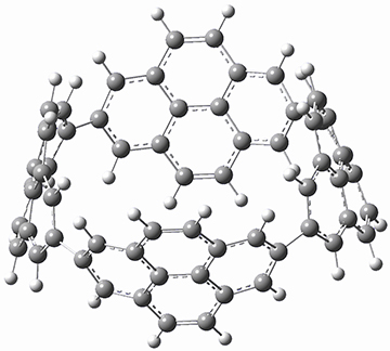
[4]Cyclo-2,7-pyrenylene

Aromaticity Steven Bachrach 09 Jul 2014 No Comments

Macrocycles composed of aromatic subunits, like polycycloparaphenylenes, are of interest as components of nanotubes and for possible interesting optical properties. Tremendous advances have occurred over the past decade in preparing these rings ; see for examples these posts. Yamago now reports on the synthesis, optical properties and structure of [4]cyclo-2,7-pyrenylene 1, made by joining four pyrene units together.1

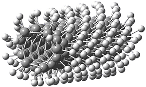
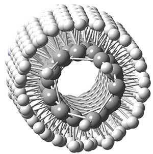
B3LYP/6-31G(d) optimization of the structure of 1 reveals a D2 geometry (Figure 1). This structure shows a very distorted pyrene unit. The strain energy of 1 is estimated as 392 kJ mol-1 (though how this was arrived at is not mentioned!), which is much larger than the strain energy of [8]-cycloparaphenylene.

Figure 1. B3LYP/6-31G(d) optimized structure of 1
This is another molecule to be sure to click on and rotate using JMol.

The nature of the HOMO and LUMO of 1 is very different than that of linear tetra-2,7-pyrene. The degenerate HOMOs and degenerate LUMOs of the linear compound have a node at the 2 and 7 positions and are localized to the terminal and central pyrene units, respectively. The HOMO and LUMO of 1 are fully delocalized. The implications of this are seen in the spectroscopy and electrochemistry of 1.

References

(1) Iwamoto, T.; Kayahara, E.; Yasuda, N.; Suzuki, T.; Yamago, S. "Synthesis, Characterization, and Properties of [4]Cyclo-2,7-pyrenylene: Effects of Cyclic Structure on the Electronic Properties of Pyrene Oligomers," Angew. Chem. Int. Ed. 2014, 53, 6430-6434, DOI: http://dx.doi.org/10.1002/anie.201403624.

InChIs

1: InChI=1S/C64H32/c1-2-34-18-50-20-36-4-3-35-19-49(17-33(1)57(35)58(34)36)51-21-37-5-7-41-25-53(26-42-8-6-38(22-51)59(37)61(41)42)55-29-45-13-15-47-31-56(32-48-16-14-46(30-55)63(45)64(47)48)54-27-43-11-9-39-23-52(50)24-40-10-12-44(28-54)62(43)60(39)40/h1-32H/b51-49-,52-50-,55-53-,56-54-
InChIKey=AWPUJIHMRRFHHU-GYSOBDCPSA-N

Structure of Citrinalin B

NMR Steven Bachrach 24 Jun 2014 4 Comments

Here is another nice example of the partnership between experiment and computation in ascertaining molecular structure. The Sarpong, Tantillo, Andersen, Berlinck, and Miller groups collaborated on the synthesis, characterization and biosynthesis of some metabolites from Penniculium strains.1 I will focus here on just the structural identification component of this paper; the synthesis and the biosynthesis are very interesting too!

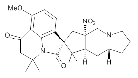
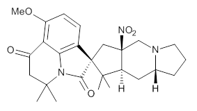
Cyclopiamine A 1 and cyclopiamine B 2 interconvert through an intermediate that allows for the epimerization at carbon bearing the nitro group.2


1


2

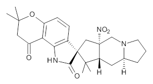
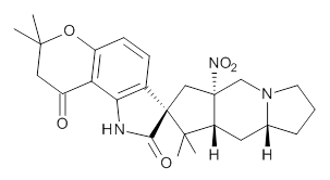
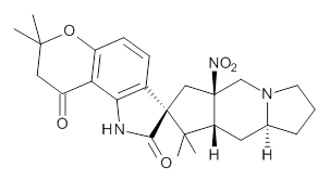
Citrinalin A 3 might also seem to undergo the same type of ring opening-ring closing reaction to produce citrinalin B. However, the original proposed structure3 of citrinalin B 4 implies an epimerization at a different carbon (at the ring fusion to the terminal 5 member ring). These authors suggested that perhaps the proper structure of citrinalin B is 5, which differs from citrinalin A only at the carbon bearing the nitro group, analogous to the relationship between 1 and 2.


3


4


5

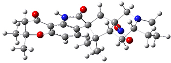
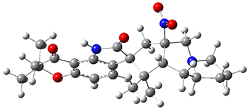
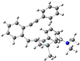
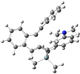
The low energy conformations of both 4 and 5 (actually the trifluoroacetic acid salts) were optimized at B3LYP/6-31+G(d,p) and the chemical shifts for both 1H and 13C were computed, Boltzmann-weighted and scaled, and then compared with the NMR spectra of authentic citrinalin B. (The lowest energy conformations of 4 and 5 are shown in Figure 1.) The corrected mean absolute deviations for the 1H and 13C chemical shift for the original structure 4 are 0.45 ppm and 2.0 ppm, respectively (with the largest outliers of 2.3 ppm for H and 9.6 ppm for C). These errors are about twice what is observed in comparing the experimental and computed 1H and 13C chemical shifts of 3. The agreement between the computed and experimental values using 5 are much improved, with mean deviations of 0.12 and 1.6ppm, and largest deviations of 0.38 ppm for 1H and 4.4 ppm for 13C. Use of Goodman’s DP4 method indicates a 100% probability that the structure of citrinalin B is 5. This prediction is confirmed by the x-ray structure.

4

5

Figure 1. B3LYP/6-31+G(d,p) optimized lowest energy conformers of 4 and 5.

References

(1) Mercado-Marin, E. V.; Garcia-Reynaga, P.; Romminger, S.; Pimenta, E. F.; Romney, D. K.; Lodewyk, M. W.; Williams, D. E.; Andersen, R. J.; Miller, S. J.; Tantillo, D. J.; Berlinck, R. G. S.; Sarpong, R. "Total synthesis and isolation of citrinalin and cyclopiamine congeners," Nature 2014, 509, 318-324, DOI: 10.1038/nature13273.

(2) Bond, R. F.; Boeyens, J. C. A.; Holzapfel, C. W.; Steyn, P. S. "Cyclopiamines A and B, novel oxindole metabolites of Penicillium cyclopium westling," J. Chem. Soc., Perkin Trans I 1979, 1751-1761, DOI: 10.1039/P19790001751.

(3) Pimenta, E. F.; Vita-Marques, A. M.; Tininis, A.; Seleghim, M. H. R.; Sette, L. D.; Veloso, K.; Ferreira, A. G.; Williams, D. E.; Patrick, B. O.; Dalisay, D. S.; Andersen, R. J.; Berlinck, R. G. S. "Use of Experimental Design for the Optimization of the Production of New Secondary Metabolites by Two Penicillium Species," J. Nat. Prod. 2010, 73, 1821-1832, DOI: 10.1021/np100470h.

InChIs

1: InChI=1S/C26H33N3O5/c1-23(2)12-17(30)20-18(34-5)9-8-16-21(20)28(23)22(31)26(16)13-25(29(32)33)14-27-10-6-7-15(27)11-19(25)24(26,3)4/h8-9,15,19H,6-7,10-14H2,1-5H3/t15-,19+,25+,26-/m1/s1
InChIKey=LUESCPPTDJZZQN-VBPSCBQFSA-N

2: InChI=1S/C26H33N3O5/c1-23(2)12-17(30)20-18(34-5)9-8-16-21(20)28(23)22(31)26(16)13-25(29(32)33)14-27-10-6-7-15(27)11-19(25)24(26,3)4/h8-9,15,19H,6-7,10-14H2,1-5H3/t15-,19+,25-,26-/m1/s1
InChIKey=LUESCPPTDJZZQN-DCWDXPITSA-N

3: InChI=1S/C25H31N3O5/c1-22(2)11-16(29)19-17(33-22)8-7-15-20(19)26-21(30)25(15)12-24(28(31)32)13-27-9-5-6-14(27)10-18(24)23(25,3)4/h7-8,14,18H,5-6,9-13H2,1-4H3,(H,26,30)/t14-,18+,24+,25-/m0/s1
InChIKey=QVBARITWSQCBBV-DTKOROOESA-N

4: InChI=1S/C25H31N3O5/c1-22(2)11-16(29)19-17(33-22)8-7-15-20(19)26-21(30)25(15)12-24(28(31)32)13-27-9-5-6-14(27)10-18(24)23(25,3)4/h7-8,14,18H,5-6,9-13H2,1-4H3,(H,26,30)/t14-,18-,24-,25+/m1/s1
InChIKey=QVBARITWSQCBBV-KUCRYEJDSA-N

5: InChI=1S/C25H31N3O5/c1-22(2)11-16(29)19-17(33-22)8-7-15-20(19)26-21(30)25(15)12-24(28(31)32)13-27-9-5-6-14(27)10-18(24)23(25,3)4/h7-8,14,18H,5-6,9-13H2,1-4H3,(H,26,30)/t14-,18+,24-,25-/m0/s1
InChIKey=QVBARITWSQCBBV-QBDGMKJCSA-N
Solvent effect on carbene spin state

carbenes Steven Bachrach 18 Jun 2014 No Comments

Carbenes remain an active area of interest for computational chemists, as seen in Chapter 5 of my book. For many carbenes, the triplet is the ground state, and that is true of diphenylcarbene 1. Can solvent play a role in the stability of carbene spin states? Surprisingly, the answer, provided in a recent paper by Sander,1 is yes!

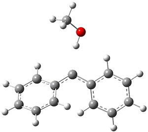
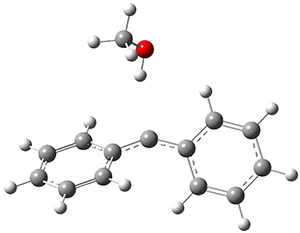
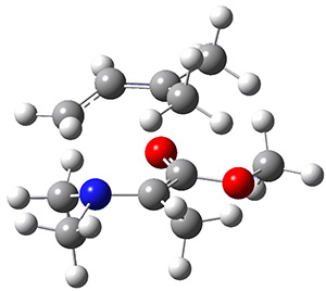
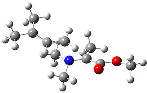
In the gas phase, the singlet-triplet gap of 1 is computed to be 5.62 kcal mol-1 at (U)B3LYP/6-311++G(d,p) (and this reduces to 5.06 at (U)B3LYP+D3/6-311++G(d,p)) with the ground state as a triplet. If a single methanol molecules is allowed to approach 1, then the complex involving the singlet has a short hydrogen bond distance of 1.97 Å but the triplet has a much longer distance of 2.33 Å. These structures are shown in Figure 1. This manifests in a dramatic change in the relative stability, with the singlet complex now 0.26 kcal mol-1 (0.44 with the D3 correction) lower in energy than the triplet.

Singlet-1:methanol

Triplet-1:methanol

Figure 1. (U)B3LYP/6-311++G(d,p) optimized geometries of the compelxes of methanol with singlet or triplet 1.

IR spectroscopy of 1 in an argon matrix doped with a small amount of methanol confirms the presence of the singlet carbene, and detailed description of the difference in the reactivities of the singlet and triplet are provided.

References

(1) Costa, P.; Sander, W. "Hydrogen Bonding Switches the Spin State of Diphenylcarbene from Triplet to Singlet," Angew. Chem. Int. Ed. 2014, 53, 5122-5125, DOI: 10.1002/anie.201400176.

InChIs

1: InChI=1S/C13H10/c1-3-7-12(8-4-1)11-13-9-5-2-6-10-13/h1-10H
InChIKey=XMGMFRIEKMMMSU-UHFFFAOYSA-N

Structure of dihydroxycarbene

carbenes Steven Bachrach 09 Jun 2014 No Comments

Dihdroxycarbene was the subject of a post a few years ago relating to how this carbene does not undergo tunneling,1 while related hydroxycarbene do undergo a tunneling rearrangement.

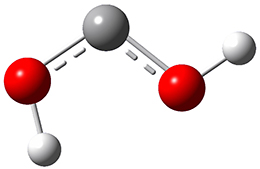
Now we have a gas-phase microwave determination of the trans,cis isomer of dihydroxycarbene.2 The computed CCSD(T)/cc-pCVQZ structure is shown in Figure 1. What is truly remarkable here is the amazing agreement between the experimental and computed structure – as seen in Table 1.The bond distance are in agreement within 0.001 Å and the bond angles agree within 0.3°! Just further evidence of the quality one can expect from high-level computations. And computing this structure was certainly far easier than the experiments!

Figure 1. CCSD(T)/cc-pCVQZ optimized geometry of dihydroxycarbene.

Table 1. Experimental and computed (CCSD(T)/cc-pCVQZ) geometric parameters of dihydroxycarbene.a


Expt.

Comp.

C-O

1.335

1.336

C-O

1.309

1.309

O-Htrans

0.961

0.960

O-Hcis

0.976

0.975

O-C-O

107.30

107.25

C-O-H­­trans

106.8

106.8

C-O-H­­cis

110.7

110.4


aDistances in Å and angles in deg.

References

(1) Schreiner, P. R.; Reisenauer, H. P. "Spectroscopic Identification of Dihydroxycarbene," Angew. Chem. Int. Ed. 2008, 47, 7071-7074, DOI: 10.1002/anie.200802105.

(2) Womack, C. C.; Crabtree, K. N.; McCaslin, L.; Martinez, O.; Field, R. W.; Stanton, J. F.; McCarthy, M. C. "Gas-Phase Structure Determination of Dihydroxycarbene, One of the Smallest Stable Singlet Carbenes," Angew. Chem. Int. Ed. 2014, 53, 4089-4092, DOI: 10.1002/anie.201311082.

InChIs

Dihydroxycarbene: InChI=1S/CH2O2/c2-1-3/h2-3H
InChIKey=VZOMUUKAVRPMBY-UHFFFAOYSA-N

[2,2]paracyclophane – structure resolved

Uncategorized Steven Bachrach 28 May 2014 3 Comments

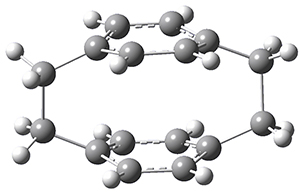
The structure of [2,2]paracyclophane 1 has been somewhat controversial for some time. Early x-ray structures indicated that the molecule was quite symmetric, D2h with the phenyl rings and the ethyl bridges eclipsed. Subsequent low-T experiments suggested a lower symmetry form D2 with a twist that relieves some of the unfavorable eclipsing interactions in the ethano bridges. High-level computations by Grimme1 and then some by myself2 indicated that the D2 structure is the lowest energy conformation, with however a low barrier through the D2h structure.

The suggestion of the D2 minimum was vehemently criticized by Dodziuk, et al. on the basis of NMR analysis.3

Now, a low temperature x-ray experiment of 1 brings clarity to the situation.4 (The introduction provides a nice summary of the previous 70 year history regarding the structure of 1.) At temperatures below 45 K, 1 is found as a single structure of D2 symmetry (with space group P4n2). The structure is shown in Figure 1. A phase change occurs at about 45 K, and above 60 K the crystal has P42/mnm symmetry. The structure of 1 at the high temperature appears as D2h with somewhat broader thermal motion of the ethano carbons than the phenyl carbons. The low T structure is in excellent accord with the previous theoretical studies, and the phase transition helps bring into accord all of the previous x-ray crystallographic work.

Figure 1. X-ray structure at 15K of 1.

References

(1) Grimme, S. "On the Importance of Electron Correlation Effects for the π-π Interactions in Cyclophanes," Chemistry Eur. J. 2004, 10, 3423-3429, DOI: 10.1002/chem.200400091.

(2) Bachrach, S. M. "DFT Study of [2.2]-, [3.3]-, and [4.4]Paracyclophanes: Strain Energy, Conformations, and Rotational Barriers," J. Phys. Chem. A 2011, 115, 2396-2401, DOI: 10.1021/jp111523u.

(3) Dodziuk, H.; Szymański, S.; Jaźwiński, J.; Ostrowski, M.; Demissie, T. B.; Ruud, K.; Kuś, P.; Hopf, H.; Lin, S.-T. "Structure and NMR Spectra of Some [2.2]Paracyclophanes. The Dilemma of [2.2]Paracyclophane Symmetry," J. Phys. Chem. A 2011, 115, 10638-10649, DOI: 10.1021/jp205693a.

(4) Wolf, H.; Leusser, D.; R. V. Jørgensen, M.; Herbst-Irmer, R.; Chen, Y.-S.; Scheidt, E.-W.; Scherer, W.; Iversen, B. B.; Stalke, D. "Phase Transition of [2,2]-Paracyclophane – An End to an Apparently Endless Story," Chem. Eur. J. 2014, 20, 7048–7053, DOI: 10.1002/chem.201304972.

InChIs

1: InChI=1S/C16H16/c1-2-14-4-3-13(1)9-10-15-5-7-16(8-6-15)12-11-14/h1-8H,9-12H2
InChIKey=OOLUVSIJOMLOCB-UHFFFAOYSA-N

Polytwistane!

Schreiner &twistane Steven Bachrach 20 May 2014 3 Comments

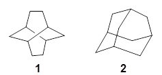
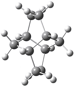
Twistane 1 is a more strained isomer of adamantane 2. The structure of 1 is shown in Figure 1.

1

Figure 1. B3LYP/6-31G(d) optimized structure of 1.

Adamantane is the core structure of diamond, which can be made by appending isobutene groups onto adamantane. In an analogous fashion, twistane can be extended in a linear way by appending ethano groups in a 1,4-bridge. Allen, Schreiner, Trauner and co-workers have examined this “polytwistane” using computational techniques.1 They examined a (CH)236 core fragment of polytwistane, with the dangling valences at the edges filled by appending hydrogens, giving a C236H242 compound. This compound was optimized at B3LYP/6-31G(d) and shown in Figure 2a. (Note that I have zoomed in on the structure, but by activating Jmol – click on the figure – you can view the entire compound.) A fascinating feature of polytwistane is its helical structure, which can be readily seen in Figure 2b. A view down the length of this compound, Figure 2c, displays the opening of this helical cylinder; this is a carbon nanotube with an inner diameter of 2.6 Å.

(a)


(b)


(c)

Figure 2. B3LYP/6-31G(d) structure of the C236H242 twistane. (a) A zoomed in look at the structure. This structure links to the Jmol applet allowing interactive viewing of the molecule – you should try this! (b) a side view clearly showing its helical nature. (c) A view down the twistane showing the nanotube structure.

Though the molecule looks quite symmetric, each carbon is involved in three C-C bonds, and each is of slightly different length. The authors go through considerable detail about addressing the symmetry and proper helical coordinates of polytwistane. They also estimate a strain energy of about 1.6 kcal mol-1 per CH unit. This modest strain, they believe, suggests that polytwistanes might be reasonable synthetic targets.

References

(1) Barua, S. R.; Quanz, H.; Olbrich, M.; Schreiner, P. R.; Trauner, D.; Allen, W. D. "Polytwistane," Chem. Eur. J. 2014, 20, 1638-1645, DOI: 10.1002/chem.201303081.

InChIs

1: InChI=1S/C10H16/c1-2-8-6-9-3-4-10(8)5-7(1)9/h7-10H,1-6H2
InChIKey=AEVSQVUUXPSWPL-UHFFFAOYSA-N

2: InChI=1S/C10H16/c1-7-2-9-4-8(1)5-10(3-7)6-9/h7-10H,1-6H2
InChIKey=ORILYTVJVMAKLC-UHFFFAOYSA-N

An approach towards identifying dynamic effect without trajectories

Dynamics Steven Bachrach 12 May 2014 No Comments

Demonstrating the occurrence of non-statistical dynamics generally has been accomplished through trajectory studies. These trajectory studies are often quite computationally demanding, requiring many trajectories, often of long duration, with molecules that are typically not small! Schmittel and co-workers present a case where their evidence for non-statistical dynamics rests not on trajectory studies but a combination of experimental product distributions and free energy of activation computations.1

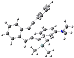
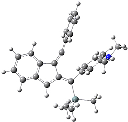
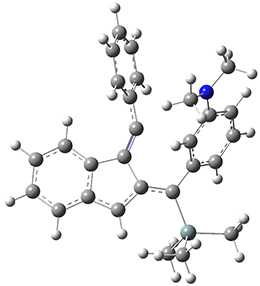
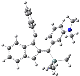
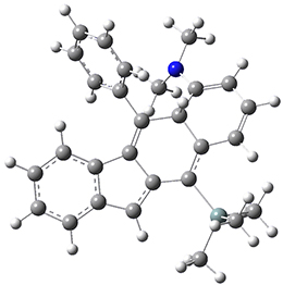
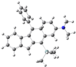
For the Schmittel C2-C6 cyclization taking 1 into 5¸Schmittel has located no concerted transition state, but rather two different transition states 2 and 2’, leading to a common intermediate diradical 3. Then there are two different transition states 4 and 4’ leading to the two regioisomeric products 5 and 5’. The BLYP/6-31G* structures and relative free energies are shown in Figure 1.

1
0.0

2
19.4

2’
20.2

3
13.3

4
16.5

4’
15.4

5
-6.3

5’
-14.3

Figure 1. BLYP/6-31G*geometries and relative free energies (kcal mol-1) of the critical points along the reaction 15.

If transition state theory (TST) holds here, the rate limiting step is the first set of transition states, and the product distribution should be dictated by the second set of transition states. Since 4’ is lower in energy than 4, TST predicts that 5’ should be the major product. However, the experiments show that the ratio 5:5’ ranges from 1.48 at 30 °C to 1.65 at 60 °C, with the ratio decreasing a bit at higher temperatures still.

Examination of the potential energy surfaces in the neighborhoods of the transition states and the intermediate show a couple of interesting features. First, there is a large barrier separating 2 and 2’ and this precludes the concerted pathway. Second, the minimum energy path forward from 2 requires a sharp turn to proceed to the intermediate 3. Schmittel suggests that this surface supports the notion of some direct reaction paths from 2 avoiding the intermediate 3 and directly over transition state 4’. Schmittel offers a simple formula for predicting the percentage of the products formed from a non-statistical pathway:

XNSQ1 + XSQ2 = Qexp

where XNS is the mole fraction following non-statistical pathways and XS is the fraction following a statistical pathway and Qexp is the experimental mole ratio and Q1 is the partitioning at the first set of TSs and Q2 is the partitioning at the second set of TSs. While this approach is certainly much simpler than performing molecular dynamics, it does require experimental values. According to this model, the above reaction follows non-statistical dynamics about 75% of the time.

References

(1) Samanta, D.; Rana, A.; Schmittel, M. "Quantification of Nonstatistical Dynamics in an Intramolecular Diels–Alder Cyclization without Trajectory Computation," J. Org. Chem. 2014, 79, 2368-2376, DOI: 10.1021/jo500035b.

InChIs

1: InChI=1S/C28H29NSi/c1-29(2)27-17-11-16-26(22-27)28(30(3,4)5)21-20-25-15-10-9-14-24(25)19-18-23-12-7-6-8-13-23/h6-17,20,22H,1-5H3
InChIKey=CKQXQJAGCGSOOP-UHFFFAOYSA-N

5: InChI=1S/C28H29NSi/c1-29(2)24-17-11-16-22-27(24)25(19-12-7-6-8-13-19)26-21-15-10-9-14-20(21)18-23(26)28(22)30(3,4)5/h6-17H,18H2,1-5H3
InChIKey=OFALZDSJOVLMHZ-UHFFFAOYSA-N

5′: InChI=1S/C28H29NSi/c1-29(2)21-15-16-23-24(18-21)28(30(3,4)5)25-17-20-13-9-10-14-22(20)27(25)26(23)19-11-7-6-8-12-19/h6-16,18H,17H2,1-5H3
InChIKey=UQRNTADMKHTLNR-UHFFFAOYSA-N

The smallest catenane?

Schaefer Steven Bachrach 06 May 2014 No Comments

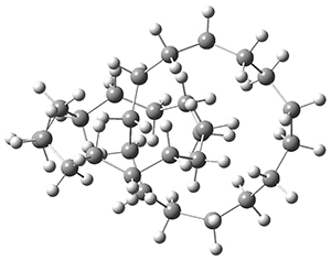
“How small can a catenane be?” This question is asked by Schaefer, Allinger and colleagues and answered (well, almost answered) using computations.1 Catenanes are linked rings. The catenanes examined here are two linked saturated hydrocarbon rings, each of the same size. The rings examined have 11 to 18 carbon atoms. The geometries were optimized with D2 symmetry, where either the closest approach between the two rings are two carbon atoms or the midpoint of two C-C bonds. The former turn out to be lower in energy. Geometries were optimized with MP2, B3LYP, BP86 or M06-2X with the DZP++ basis set. There is little geometric dependence on computational method. The optimized geometry of the catenane with 14 carbons is shown in Figure 1.

Figure 1. Optimized geometry of the 14-carbon catenane. (Be sure to click on this structure to view the molecule in 3-D; you will have to allow Jmol to download and run!)

To cut to the chase, as the rings get smaller they observe a lengthening of the C-C bonds at the intersection. With the 14-carbon catenane they observe a significant increase in the bond length near the intersection, suggesting a dramatic instability. This is also seen in the change in the energy per C as the rings get smaller; a large increase in energy per C is seen at the transition from 14 to 13 carbons. This all points toward the 14-carbon catenane as the smallest one that might be stable.

(I thank Prof. Schaefer and colleagues for providing me with the coordinates of the 14-carbon catenane.)

References

(1) Feng, X.; Gu, J.; Chen, Q.; Lii, J.-H.; Allinger, N. L.; Xie, Y.; Schaefer, H. F. "How Small Can a Catenane Be?," J. Chem. Theor. Comput. 2014, 10, 1511-1517, DOI: 10.1021/ct400926p

Dynamic effects in [1,2]- and [2,3]-sigmatropic rearrangements

Dynamics &Singleton Steven Bachrach 29 Apr 2014 No Comments

While the [2-3]-sigmatropic rearrangement is well known and understood as allowed under the Woodward-Hoffmann rules, [1,2]-sigmatropic are much more rare, perhaps because they are forbidden by the same orbital symmetry arguments. It is perhaps surprising that these two rearrangements may sometimes be found in competition. Singleton has applied many of his tried-and-true techniques, namely, careful normal abundance kinetic isotope effect (KIE) analysis and molecular dynamics computations, to this problem.1

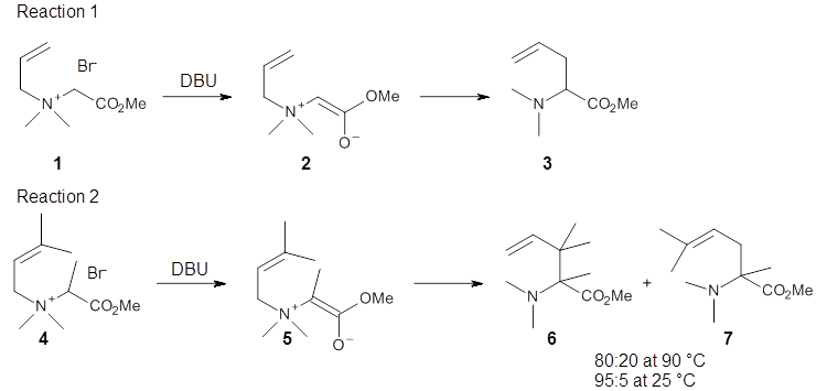
Reaction 1 takes place exclusively through a [2,3]-rearrangement; the principle evidence is the lack of any crossover reaction. However, the slightly more substituted analogue shown in Reaction 2 gives rise to two products: that obtained from a [2,3]-rearrangement 6 and that obtained from a [1,2]-rearrangement 7.

The KIE for the rearrangement of 2 is large for the carbon breaking the bond with nitrogen, while it is small at the carbons that are forming the new bond. This becomes a metric for judging the transition state obtained with computations. With the computed TS and canonical variational transition state theory (VTST) including small curvature tunneling, the KIE can be computed from a computed structures and frequencies. This imposes a range of reasonable distances for the forming C-C bond of 2.6-2.9 Å – much longer that a typical distance in the TS of similar pericyclic reactions.

Crossover experiments for Reaction 2 are understood in terms of a reaction model whereby some fraction of the reactants undergo a concerted rearrangement to form 6, and 7 is formed by first breaking the C-N bond, forming two radicals, that either recombine right away or form isolated radicals that then collapse to product.

The interesting twist here is that one would expect two different transition states, one for the concerted process 8 and one to cleave the bond 9. Both do exist and are shown in Figure 1. However, VTST predicts that the concerted process should be 25-50 times faster than cleavage, and that does not match up with experiments. Amazingly, molecular dynamics trajectories started from the concerted TS 8 leads to cleavage about 20% of the time using UMO6-2X with a variety of basis sets. Thus, as Singleton has noted many times before, a single TS is crossed that leads to two different products! An argument based on entropy helps explain why the second (cleavage) pathway is viable.

8

9

Figure 1. UMO6-2x/6-31G* optimized structures of TS 8 and 9.

References

(1) Biswas, B.; Collins, S. C.; Singleton, D. A. "Dynamics and a Unified Understanding of Competitive [2,3]- and [1,2]-Sigmatropic Rearrangements Based on a Study of Ammonium Ylides," J. Am. Chem. Soc. 2014, 136, 3740-3743, DOI: 10.1021/ja4128289.

Dispersion – application to cellular membranes

Schreiner Steven Bachrach 22 Apr 2014 1 Comment

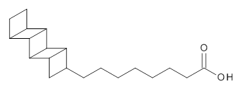
Schreiner provides another beautiful example of the important role that dispersion plays, this time in a biological system.1 The microbe Candidatus Brocadia Anammoxidans oxidizes ammonia with nitrite. This unusual process must be done anaerobically and without allowing toxic side products, like hydrazine to migrate into the cellular environment. So this cell has a very dense membrane surrounding the enzymes that perform the oxidation. This dense membrane is home to some very unusual lipids, such as 1. These lipids contain the ladderane core, a highly strained unit. Schreiner hypothesized that these ladderane groups might pack very well and very tightly due to dispersion.


1

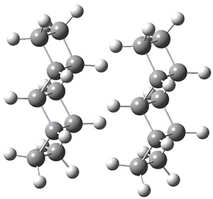
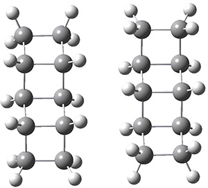
The geometries of the [2]- through [5]-ladderanes and their dimers were optimized at MP2/aug-cc-pVDZ, and the binding energies corrected for larger basis sets and higher correlation effects. The dimers were oriented in their face-to-face orientation (parallel-displaced dimer, PDD) or edge-to-edge (side-on dimer, SD). Figure 1 shows the optimized structures of the two dimeric forms of [4]-ladderane.

[4]-PDD

[4]-SD

Figure 1. MP2/aug-cc-pVDZ optimized geometries of the dimers of [4]-ladderane in the PDD and SD orientations.

The binding energies of the ladderane dimers, using the extrapolated energies and at B3LYP-D3/6-311+G(d,p), are listed in Table 1. (The performance of the B3LYP-D3 functional is excellent, by the way.)The binding is quite appreciable, greater than 6 kcal mol-1 for both the [4]- and [5]-ladderanes. Interestingly, these binding energies far exceed the binding energies of similarly long alkanes. So, very long alkyl lipid chains would be needed to duplicate the strong binding. Nature appears to have devised a rather remarkable solution to its cellular isolation problem!


Eextrapolated

EB3LYP-D3

[2]-PDD

-27.

-3.2

[3]-PDD

-4.2

-4.1

[4]-PDD

-5.5

-5.3

[5]-PDD

-6.6

-6.5

[2]-SD

-3.3

-3.1

[3}-SD

-4.1

-4.4

[4]-SD

-6.5

-5.7

[5]-SD

-7.5

-7.0

References

(1) Wagner, J. P.; Schreiner, P. R. "Nature Utilizes Unusual High London Dispersion
Interactions for Compact Membranes Composed of Molecular Ladders," J. Chem. Theor. Comput. 2014, 10, 1353-1358, DOI: 10.1021/ct5000499.

InChIs

1: InChI=InChI=1S/C20H30O2/c21-15(22)7-5-3-1-2-4-6-11-10-14-16(11)20-18-13-9-8-12(13)17(18)19(14)20/h11-14,16-20H,1-10H2,(H,21,22)/t11-,12-,13+,14-,16+,17+,18-,19-,20+/m0/s1
InChIKey=ZKKJRZDMLYQUNK-QIPWGTBCSA-N

[2]-ladderane: InChI=1S/C6H10/c1-2-6-4-3-5(1)6/h5-6H,1-4H2/t5-,6+
InChIKey=YZLCEXRVQZNGEK-OLQVQODUSA-N

[3]-ladderane: InChI=1S/C8H12/c1-2-6-5(1)7-3-4-8(6)7/h5-8H,1-4H2/t5-,6+,7+,8-
InChIKey=YTZCZYFFHKYOBJ-SOSBWXJGSA-N

[4]-ladderane: InChI=1S/C10H14/c1-2-6-5(1)9-7-3-4-8(7)10(6)9/h5-10H,1-4H2/t5-,6+,7+,8-,9-,10+
InChIKey=VZHFDSXKIJOCAY-UXAOAXNSSA-N

[5]-ladderane: InChI=1S/C12H16/c1-2-6-5(1)9-10(6)12-8-4-3-7(8)11(9)12/h5-12H,1-4H2/t5-,6+,7-,8+,9+,10-,11-,12+
InChIKey=CWUAAECPDWVJSU-SBBGGFAWSA-N

« Previous PageNext Page »