Archive for the 'DFT' Category

π-π Stacking

I did not present π-π stacking in the book, but I think if I ever do a second edition, I will include a discussion of it. I’m not sure quite where it would fit in given the current structure of the book (I discuss DNA bases and base pairs in the context of solvation in Chapter 6), but the paper I will discuss next gives me some idea – π-π stacking is a sensitive test of the quality of computational methods and this could be part of Chapter 1 as a discussion of the failings of methods, especially DFT.

Swart and Bickelhaupt have examined a series of π-π stacked pairs, evaluating them regarding how DFT performs.1 Their first example is the benzene dimer (Table 1). At CCSD(T) the dimer binding energy is 1.7 kcal mol-1 and the rings are 3.9 Å apart. LDA, KT1 (yet another newly minted functional2,3), and BHandH get the separation and binding energy reasonably well. PW91 gets the distance too big and underestimates the binding energy. But most important is that the other (more traditional) functionals indicate that the PES is entirely repulsive! This is a manifestation of many functionals’ inability to properly account for dispersion.

Table 1. Optimized separation distance (rmin, Å) and binding energy (kcal mol-1)
of the benzene dimer using the TZ2P basis set.1


Method

rmin

ΔE

CCSD(T)

3.9

-1.70

LDA

3.8

-1.33

KT1

3.8

-1.58

BHandH

3.9

-0.89

PW91

5.0

-0.45

BLYP

repulsive

BP86

repulsive

OLYP

repulsive

B3LYP

repulsive


Next, they compare 14 different orientations of stacked dimmers of cytosine. The energies of these dimmers were computed using again a variety of functionals and compared to MP2/CBS energies with a correction for CCSD(T). The mean absolute deviations (MAD) for the energies using the various functionals are listed in Table 2. Again, LDA and KT1 perform quite well, but most functionals do quite poorly.

Table 2. Mean absolute deviations of the energies of 14 cytosine
stacked dimer structures compared to their MP2 energies.


Method

MAD

LDA

0.38

KT1

0.47

BHandH

0.52

PW91

6.04

BLYP

9.52

BP86

8.75

OLYP

14.80

B3LYP

8.24


Similar results are also demonstrated for stacked DNA bases and also stacked base pairs. These authors conclude that the KT1 functional appears suitable for treating π-π stacking. One should also consider some of the new functionals from the Truhlar group,4-6 which unfortunately are not included in this study.

References

(1) Swart, M.; van der Wijst, T.; Fonseca, C.; Bickelhaput, F. M., "π-π Stacking Tackled with Density Functional Theory," J. Mol. Model. 2007, 13, 1245-1257, DOI: 10.1007/s00894-007-0239-y.

(2) Keal, T. W.; Tozer, D. J., "The Exchange-Correlation Potential in Kohn–Sham Nuclear Magnetic Resonance Shielding Calculations," J. Chem. Phys. 2003, 119, 3015-3024, DOI: 10.1063/1.1590634

(3) Keal, T. W.; Tozer, D. J., "A Semiempirical Generalized Gradient Approximation Exchange-Correlation Functional," J. Chem. Phys. 2004, 121, 5654-5660, DOI: 10.1063/1.1784777.

(4) Zhao, Y.; Truhlar, D. G., "A Density Functional That Accounts for Medium-Range Correlation Energies in Organic Chemistry," Org. Lett. 2006, 8, 5753-5755, DOI: 10.1021/ol062318n

(5) Zhao, Y.; Schultz, N. E.; Truhlar, D. G., "Design of Density Functionals by Combining the Method of Constraint Satisfaction with Parametrization for Thermochemistry, Thermochemical Kinetics, and Noncovalent Interactions," J. Chem. Theory Comput., 2006, 2, 364-382, DOI: 10.1021/ct0502763.

(6) Zhao, Y.; Truhlar, D. G., "Assessment of Model Chemistries for Noncovalent Interactions," J. Chem. Theory Comput. 2006, 2, 1009-1018, DOI: 10.1021/ct060044j
DFT Steven Bachrach 26 Nov 2007 No Comments

Amino acid acidity

Poutsma has followed up on the work he reported earlier in collaboration with Kass concerning the gas-phase acidity of the amino acids.1 Their previous work reported on cysteine,2 with the unusual result that the thiol group is more acidic than the carboxylic acid group. (I blogged on this a previous post.) Now, he reports the experimental and DFT acidities of all 20 amino acids, shown in Table 1. The experiments were done using the kinetic method in a quadrupole ion trap with electrospray ionization. The computations were performed at B3LYP/6-311++G**//B3LYP/6-31+G*, following some MM searching to identify low-lying conformations. The computed acidities were obtained relative to acetic acid, i.e. R-CH2COOH + OAc → R-CH2COO +HOAc.

Table 1. Relative acidities (kJ mol-1) of the amino acids1


Exp

DFT

Gly (1434 ± 9)

Gly (1434)

Pro (1431 ± 9)

Ala (1432)

Val (1431 ± 8 )

Pro (1430)

Ala (1430 ± 8 )

Val (1430)

Ile (1423 ± 8 )

Leu (1428)

Trp (1421 ± 9)

Ile (1426)

Leu (1419 ± 10)

Trp (1422)

Phe (1418 ± 18)

Tyr (1419)

Lys (1416 ± 7)

Phe (1417)

Tyr (1413 ± 11)

Lys (1415)

Met (1407 ± 9)

Met (1412)

Cys (1395 ± 9)

Thr (1397)

Ser (1391 ± 22)

Cys (1396)

Thr (1388 ± 10)

Ser (1392)

Asn (1385 ± 9)

Arg (1387)

Gln (1385 ± 11)

Asn (1384)

Arg (1381 ± 9)

Gln (1378)

His (1375 ± 8 )

His (1374)

Glu (1348 ± 2)

Glu (1349)

Asp (1345 ± 14)

Asp (1345)


The computed values are in very good agreement with the experimental values. The amino acids are ordered in increasing acidity in Table 1. The order predicted by experiment and DFT are quite close, and the disagreements are well within the error bar of the experiment.

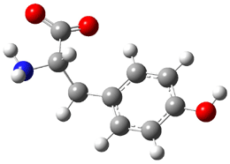
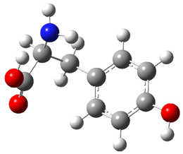
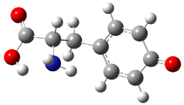
Similar to the result for cysteine, tyrosine also displays unusual acidity. The alcohol proton is more acidic than the carboxylic acid proton. The structures of tyrosine, and its two conjugate
bases, one from loss of the phenolic proton and the other from loss of the carboxylic acid proton are shown in Figure 1. The stability of the tyrosine conjugate base from loss of the phenolic
hydrogen arises from both the stability of phenoxide and the internal hydrogen bond from the carboxylic acid proton to the amine. This is different that in the cysteine case, the thiolate anion is stabilized by an internal hydrogen bond from the carboylic acid group (see Figure 2c here).

tyrosine

Tyrosine conjugate
base
(loss of phenolic hydrogen)

Tyrosine conjugate
base
(loss of carboxylate hydrogen)

Figure 1. B3LYP/6-31G* optimized structures of tyrosine and its conjugate bases.1

References

(1) Jones, C. M.; Bernier, M.; Carson, E.; Colyer, K. E.; Metz, R.; Pawlow, A.; Wischow, E. D.; Webb, I.; Andriole, E. J.; Poutsma, J. C., "Gas-Phase Acidities of the 20 Protein Amino Acids," Int. J. Mass Spectrom. 2007, 267, 54-62, DOI: 10.1016/j.ijms.2007.02.018.

(2) Tian, Z.; Pawlow, A.; Poutsma, J. C.; Kass, S. R., "Are Carboxyl Groups the Most Acidic Sites in Amino Acids? Gas-Phase Acidity, H/D Exchange Experiments, and Computations on Cysteine and Its Conjugate Base," J. Am. Chem. Soc. 2007, 129, 5403-5407, DOI: 10.1021/ja0666194.

InChI

Tyrosine: InChI=1/C9H11NO3/c10-8(9(12)13)5-6-1-3-7(11)4-2-6/h1-4,8,11H,5,10H2,(H,12,13)/t8-/m0/s1

Acidity &amino acids &DFT Steven Bachrach 12 Nov 2007 No Comments

Bergman cyclization and [10]annulenes

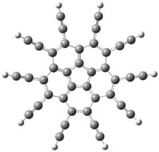
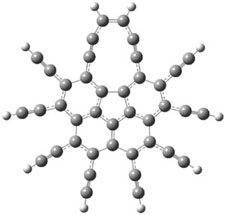
In their continuing efforts to build novel aromatic systems, Siegel and Baldridge report the preparation of the decapropyl analogue of the per-ethynylated corrannulene 1.1 They were hoping that this might cyclize to the bowl 2. It is however stable up to 100 °C, however, the analogue 3 was obtained in the initial preparation of decapropyl-1.

The B3LYP/cc-pVDZ optimized structures of 1 and 3 are shown in Figure 1. 1 is bowl-shaped, reflecting the property of corranulene, but interestingly 3 is planar. The geometry of the {10]annulene is interesting as it is more consistent with the alkynyl resonance form B.

1

3

Figure 1. B3LYP/cc-pVDZ optimized structures of 1 and 3.1

Siegel and Baldridge speculate that the conversion of 1 → 3 occurs by first undergoing the Bergman cyclization to give 4, which then opens to give 3. Unfortunately, they did not compute the activation barrier for this process. They do suggest that further cyclization to give the hoped for 2 might be precluded by the long distances between radical center and neighboring alkynes in 4, but the radicals are too protected to allowing trapping by the solvent, allowing for the formation of 3.

References

(1) Hayama, T.; Wu, Y. T.; Linden, A.; Baldridge, K. K.; Siegel, J. S., "Synthesis, Structure, and Isomerization of Decapentynylcorannulene: Enediyne Cyclization/Interconversion of C40R10 Isomers," J. Am. Chem. Soc., 2007, 129, 12612-12613 DOI: 10.1021/ja074403b.

InChIs

1: InChI=1/C40H10/c1-11-21-22(12-2)32-25(15-5)26(16-6)34-29(19-9)30(20-10)35-28(18-8)27(17-7)33-24(14-4)23(13-3)31(21)36-37(32)39(34)40(35)38(33)36/h1-10H

2: InChI=1/C40H10/c1-2-12-14-5-6-16-18-9-10-20-19-8-7-17-15-4-3-13-11(1)21-22(12)32-24(14)26(16)34-29(18)30(20)35-28(19)27(17)33-25(15)23(13)31(21)36-37(32)39(34)40(35)38(33)36/h1-10H

3: InChI=1/C40H12/c1-9-23-25(11-3)33-27(13-5)29(15-7)35-30(16-8)28(14-6)34-26(12-4)24(10-2)32-22-20-18-17-19-21-31(23)36-37(32)39(34)40(35)38(33)36/h1-8,17-18,31-32H/b18-17-

annulenes &Bergman cyclization &DFT &polycyclic aromatics Steven Bachrach 05 Nov 2007 No Comments

Predicting NMR chemical shifts of penam β-lactams

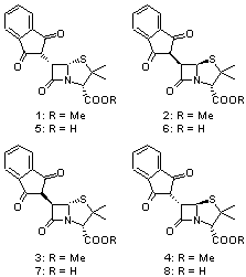
Cramer and Hoye have applied DFT computations to the predictions of both protons and carbon NMR chemical shifts in penam β-lactams1 using the procedure previously described in my blog post Predicting NMR chemical shifts. They examined the compounds 1-8 by optimizing low energy conformers at B3LYP/6-31G(d) with IEFPCM (solvent=chloroform). The chemical shifts were then computed using these geometries with the larger 6-311+G(2d,p) basis set and four different functionals: B3LYP, PBE1 and the two specific functionals designed to produce proton and carbon chemical shifts: WP04 and WC04.

A number of interesting results are reported. First, all three functionals do a fine job in predicting the proton chemical shifts of 1-8, with WP04 slightly better than the other two.On the other hand, all three methods fail to predict the carbon chemical shifts of 1-3, though B3LYP and PBE1 do correctly identify 5-8. The failure of WC04 is surprising, especially since dimethyl disulfide was used in the training set. They also noted that WP04 using just the minimum energy conformation (as opposed to a Boltzmann averaged chemical shift sampled from many low energy conformers) did correctly identify lactams 1-4. This is helped by the fact that the lowest energy conformer constituted anywhere form 37% to 68% of the energy-weighted population.

References


(1) Wiitala, K. W.; Cramer, C. J.; Hoye, T. R., “Comparison of various density functional methods for distinguishing stereoisomers based on computed 1H or 13C NMR chemical shifts using diastereomeric penam ?-lactams as a test set,” Mag. Reson. Chem., 2007, 45, 819-829, DOI: 10.1002/mrc.2045.

InChIs

1: InChI=1/C18H17NO5S/c1-18(2)14(17(23)24-3)19-15(22)11(16(19)25-18)10-12(20)8-6-4-5-7-9(8)13(10)21/h4-7,10-11,14,16H,1-3H3/t11-,14+,16+/m0/s1

5: InChI=1/C17H15NO5S/c1-17(2)13(16(22)23)18-14(21)10(15(18)24-17)9-11(19)7-5-3-4-6-8(7)12(9)20/h3-6,9-10,13,15H,1-2H3,(H,22,23)/t10-,13+,15+/m0/s1

Cramer &DFT &NMR Steven Bachrach 22 Oct 2007 No Comments

The solvent’s role in optical rotation

Bertran and Wipf have examined the role of solvent organization about a chiral molecule in producing the optical activity.1 They generated 1000 configurations of benzene arrayed about methyloxirane from a Monte Carlo simulation. Each configuration was then constructed by keeping every benzene molecules within 0.5 nm from the center-of-mass of methyloxirane, usually 8-10 solvent molecules. The optical rotation was then computed at four wavelengths using TDDFT at BP86/SVP. (The authors note that though the Gaussian group recommends B3LYP/aug-ccpVDZ,2-4 using the non-hybrid functional allows the use of resolution-of–the-identity5 techniques that make the computations about six orders of magnitude faster – of critical importance given the size of the clusters and the sheer number of them!) Optical rotation is then obtained by averaging over the ensemble.

The computed optical rotations disagree with the experiment by about 50% in magnitude but have the correct sign across the four different wavelengths. Use of the COSMO model (implicit solvent) provides the wrong sign at short wavelengths. But perhaps most interesting is that the computed optical activity of the solvent molecules in the configuration about the solute, but without including methyloxirane, is nearly identical to that of the whole cluster! In other words, the optical activity is due to the dissymmetric distribution of the solvent molecules about the chiral molecule, not the chiral molecule itself! It is the imprint of the chiral molecule on the solvent ordering that accounts for nearly all of the optical activity.

References

(1) Mukhopadhyay, P.; Zuber, G.; Wipf, P.; Beratan, D. N., "Contribution of a Solute’s
Chiral Solvent Imprint to Optical Rotation," Angew. Chem. Int. Ed. 2007,
46, 6450-6452, DOI: 10.1002/anie.200702273

(2) Stephens, P. J.; McCann, D. M.; Cheeseman, J. R.; Frisch, M. J., "Determination of
absolute configurations of chiral molecules using ab initio time-dependent Density Functional Theory calculations of optical rotation: How reliable are absolute configurations obtained for molecules with small rotations?," Chirality 2005, 17, S52-S64, DOI: 10.1002/chir.20109.

(3) Stephens, P. J.; Devlin, F. J.; Cheeseman, J. R.; Frisch, M. J., "Calculation of Optical Rotation Using Density Functional Theory," J. Phys. Chem. A 2001, 105, 5356-5371, DOI: 10.1021/jp0105138.

(4) Stephens, P. J.; McCann, D. M.; Devlin, F. J.; Flood, T. C.; Butkus, E.; Stoncius,
S.; Cheeseman, J. R., "Determination of Molecular Structure Using Vibrational Circular Dichroism Spectroscopy: The Keto-lactone Product of Baeyer-Villiger Oxidation of (+)-(1R,5S)-Bicyclo[3.3.1]nonane-2,7-dione," J. Org. Chem. 2005, 70, 3903-3913, DOI: 10.1021/jo047906y.

(5) Eichkorn, K.; Treutler, O.; Ohm, H.; Haser, M.; Ahlrichs, R., "Auxiliary Basis Sets to Approximate Coulomb Potentials," Chem. Phys. Lett. 1995, 240, 283-289, DOI: 10.1016/0009-2614(95)00621-A.

DFT &Optical Rotation &Solvation Steven Bachrach 08 Oct 2007 1 Comment

Computing optical activities

A growing area for the application of computational chemistry is in the structural identification of compounds. In the book, I discussed the use of computed IR spectra to identify intermediates in the photolysis of phenyl nitrene and carbine and the benzynes. In previous blogs, I have written posts (here, here and here) about recent use of computed NMR spectra to discern the structure of new natural products. With this post I discus the use of computed optical activity to determine the absolute configuration of molecules.

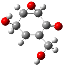
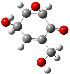
Rosini and co-workers have examined a number of oxygenated cyclohexene epoxides.to explore the use of TDDFT computed optical activity as a means for determining absolute configuration.1 In chapter 1.6.3, I discuss the work of the Gaussian team on benchmarking optical rotation and ORD. They find that B3LYP/aug-cc-pVDZ computed optical activities are in quite reasonable agreement with experiment.2-4 In this work, Rosini explores using a smaller basis set (6-31G(d)), the role of solvent, and also if computed spectra can be used to assess the absolute configuration of new molecules.

They first benchmark the B3LYP/6-31G(d) computed optical activities for a number of related cyclohexene epoxides against B3LYP/aug-cc-pVDZ and experimental values. I will begin by discussing two of their examples: (+)-chaloxone 1 (PubChem)
and (+)-epoxydon 2
(PubChem).

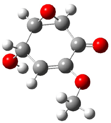
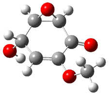
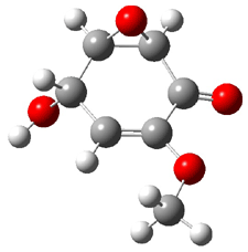
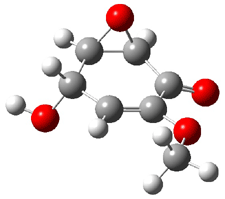
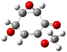
Five conformations of 1 were optimized in the gas phase and then their optical activities for the sodium D line were computed using TDDFT with both the small and larger basis set. These computations were then repeated to model the effect of solvent using PCM; the solution (methanol) B3LYP/6-31G(d) structures are shown in Figure 1.

1a

0.0

1b

2.40

1c

0.87

1d

1.02

1e

3.12


Figure 1. PCM(methanol)/B3LYP/6-31G(d) optimized structures of 1. Relative free energies of each conformer in kcal/mol.1

The optical rotation at the sodium D line was then computed with TDDFT in both gas and solution phase with the smaller and larger basis set. The values were then averaged base on a Boltzmann weighting using the computed free energies of each conformer. The optical rotation for each conformer and the average values are listed in Table 1. The experimental optical rotation is +271. The authors note that while the gas phase B3LYP/6-31G(d) average value is far off the experimental value, it does predict the correct sign, and since all of the five conformers give rise to a positive rotation, any error in the energies will not affect the sign. The computed gas phase value with the larger basis set is in better agreement with experiment. However, it is still too large, but the solution values are much better. In fact, the PCM/B3LYP/aug-cc-pVDZ value is in excellent agreement with experiment.

Table 1. Computed optical activity of the conformers of 1 in gas and solution phase.


gas

solution

conformer

6-31G(d)

aug-cc-pvDZ

6-31G(d)

aug-cc-pvDZ

1a

+264

+251

+304

+308

1b

+723

+750

+690

+707

1c

+324

+309

+398

+385

1d

+187

+201

+246

+268

1e

+741

+785

+756

+769

Averagea

+378

+333

+318

+322


aBased on a Boltzmann weighting of the population of each conformation.

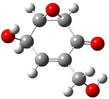
Five conformers of epoxydon 2 were also located, and the computed solution structures are shown in Figure 2. The computed optical rotations for both the gas and solution phase for these structures (and the Boltzmann weighted averages) are listed in Table 2. The experimental value for the optical rotation of 2 is +93.

2a

0.0

2b

0.32

2c

0.23

2d

0.22

2e

0.66


Figure 2. PCM(methanol)/B3LYP/6-31G(d) optimized structures of 2. Relative free energies of each conformer in kcal/mol.1

In this case, the small basis set performs very poorly. The gas phase B3LYP/6-31G(d) value
of [α]D is -16, predicting the wrong sign, let alone the wrong magnitude. Things improve with the larger basis set, which predicts a value of +57. Since conformer 2ais levorotatory and the other four are dextrorotatory, the computed relative energies are key to getting the correct prediction. This is made even more poignant with the solution results, where the PCM/B3LYP/aug-cc-pVDZ prediction is quite acceptable.

Table 2. Computed optical activity of the conformers of 2 in gas and solution phase.


gas

solution

conformer

6-31G(d)

aug-cc-pvDZ

6-31G(d)

aug-cc-pvDZ

2a

-97

-43

-85

-36

2b

+130

+210

+113

+166

2c

+14

+63

+8

+58

2d

+113

+119

+37

+71

2e

+29

+86

+19

+67

Averagea

-16

+57

+4

+61


aBased on a Boltzmann weighting of the population of each conformation.

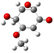
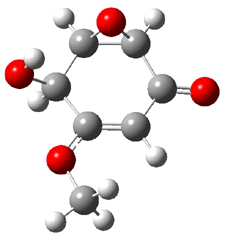
Threy conclude with two examples of application of computation to assignment of structure. I discuss here the absolute configuration of (-)-sphaeropsidone 3 (PubChem).
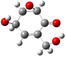
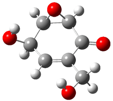
Rosini located two conformations of 3, shown in Figure 3. The computed optical rotations are listed in Table 3. The experimental value for 3 is -130. Both conformers are computed to be dextrorotatory with all computational methods. The magnitude of the computed values using the larger basis set is in nice agreement with experiment, but the sign is wrong. Rosini concludes that the absolute configuration of 3 has been misassigned.

3a

0.06

3b

0.0

Figure 3. PCM(methanol)/B3LYP/6-31G(d) optimized structures of 3. Relative free energies of each conformer in kcal/mol.1

Table 3. Computed optical activity of the conformers of 3 in gas and solution phase.


gas

solution

conformer

6-31G(d)

aug-cc-pvDZ

6-31G(d)

aug-cc-pvDZ

3a

+99

+172

+67

+135

3b

+54

+109

+20

+69

Averagea

+85

+146

+43

+101


aBased on a Boltzmann weighting of the population of each conformation.

References

(1) Mennucci, B.; Claps, M.; Evidente, A.; Rosini, C., "Absolute Configuration of Natural Cyclohexene Oxides by Time Dependent Density Functional Theory Calculation of the Optical Rotation: The Absolute Configuration of (-)-Sphaeropsidone and (-)-Episphaeropsidone Revised," J. Org. Chem. 2007, 72, 6680-6691, DOI: 10.1021/jo070806i

(2) Stephens, P. J.; Devlin, F. J.; Cheeseman, J. R.; Frisch, M. J., "Calculation of Optical Rotation Using Density Functional Theory," J. Phys. Chem. A 2001, 105, 5356-5371, DOI: 10.1021/jp0105138.

(3) Stephens, P. J.; McCann, D. M.; Cheeseman, J. R.; Frisch, M. J., "Determination of
absolute configurations of chiral molecules using ab initio time-dependent Density Functional Theory calculations of optical rotation: How reliable are absolute configurations obtained for molecules with small rotations?," Chirality 2005, 17, S52-S64, DOI: 10.1002/chir.20109.

(4) Stephens, P. J.; McCann, D. M.; Devlin, F. J.; Flood, T. C.; Butkus, E.; Stoncius,
S.; Cheeseman, J. R., "Determination of Molecular Structure Using Vibrational Circular Dichroism Spectroscopy: The Keto-lactone Product of Baeyer-Villiger Oxidation of (+)-(1R,5S)-Bicyclo[3.3.1]nonane-2,7-dione," J. Org. Chem. 2005, 70, 3903-3913, DOI: 10.1021/jo047906y.

InChI

1: InChI=1/C7H8O4/c1-10-4-2-3(8)6-7(11-6)5(4)9/h2-3,6-8H,1H3

2: InChI=1/C7H8O4/c8-2-3-1-4(9)6-7(11-6)5(3)10/h1,4,6-9H,2H2

3: InChI=1/C7H8O4/c1-10-4-2-3(8)6-7(11-6)5(4)9/h2,5-7,9H,1H3

DFT &Optical Rotation Steven Bachrach 24 Sep 2007 No Comments

Metal-assisted Cope rearrangements

Despite the fact that Wes Borden has indicated the he has written his last paper on the Cope rearrangement (see my interview with Wes at the end of Chapter 3), others remain intrigued by this reaction and continue to report on it. In a recent JACS communication, Tantillo1 examines the palladium-promoted Cope rearrangement.

The ordinary Cope rearrangement displays chameleonic character – switching from concerted to stepwise with a diradical intermediate – based on substituents. The palladium-promoted Cope is suggested to proceed through a stepwise mechanism with a zwitterionic intermediate (Scheme 1).2

Scheme 1.

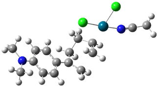
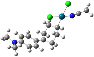
Tantillo1 has examined a variety of these rearrangements at the B3LYP/LANL2DZ level. The palladium complex is PdCl2NCMe. For all cases where R is a substituted phenyl group, the mechanism is stepwise, with the intermediate 1 sitting in a shallow well. The most stable intermediate (based on lying in the deepest well) is with the 4-dimethylaminophenyl group, and the well is 5.1 kcal mol-1 deep. The structures of the transition state (2-pNMe2) and the intermediate (1-pNMe2) are shown in Figure 1.

2-pNMe2

1-pNMe2

Figure 1. B3LYP/LANL2DZ optimized structures of 2-pNMe2 and 1-pNMe2.1

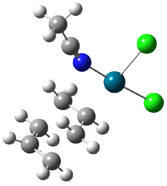
However, the well associated with 1 can be very shallow, as little as 0.4 kcal mol-1 (R = 4-trifluoroimethylphenyl and 4-nitrophenyl). This suggests that perhaps when properly substituted the intermediate might vanish and the reaction become concerted. This is in fact what happens when R is CF3, CN, or H. The transition state for the reaction with R = H is shown in Figure 2. So, this metal-assisted Cope rearrangement displays chameleonic behavior, just like the metal-free case, except that the intermediate is zwitterionic with the metal, instead of diradical in the metal-free cases.

2-H

Figure 1. B3LYP/LANL2DZ optimized structure of 2-H.1

References

(1) Siebert, M. R.; Tantillo, D. J., "Transition-State Complexation in Palladium-Promoted [3,3] Sigmatropic Shifts," J. Am. Chem. Soc. 2007, 129, 8686-8687, DOI: 10.1021/ja072159i.

(2) Overman, L. E.; Renaldo, A. E., "Catalyzed Sigmatropic Rearrangements. 10. Mechanism of the Palladium Dichloride Catalyzed Cope Rearrangement of Acyclic Dienes. A Substituent Effect Study," J. Am. Chem. Soc. 1990, 112, 3945-3949, DOI: 10.1021/ja00166a034.

Cope Rearrangement &DFT Steven Bachrach 04 Sep 2007 No Comments

Computing Accurate Energies

A couple of additional papers have pointed out systematic problems with using DFT and offer guidelines for methods that provide accurate results. These complement my previous posts on the subject Problems with DFT and Problems with DFT – an Update.

Grimme1 takes the approach of benchmarking methods and basis sets using isomerization energies, examples of which are shown in Scheme 1. Computed isomerization reaction energies are compared against experimental values or, in a few cases, against extrapolated CCSD(T) energies using cc-pVXZ (X=D-T or X=T-Q). This extrapolation technique2 is a way to estimate the complete basis set energy.

Scheme 1.

In terms of basis set, the error systematically decreases with increasing size of the basis set when the SCS-MP2 method is used to compute the energies. Surprisingly, the error is essentially constant for all the basis sets with B3LYP. The root-mean-square deviation and maximum error for the isomerization energies computed with the TZV(2df,2pd) basis set and a variety of different methods are listed in Table 1. Both CCSD(T) and SCS-MP2 provide truly excellent results. Since the later method is much more computationally efficient that the former, Grimme argues that this is really the method of choice for accurate energies. DFT methods vary in their performance, with no discernable trend based on what type of DFT it is (i.e. meta-GGA, hybrid GGA, or hybrid meta-GGA). Of no surprise, based on lots of recent studies (including those blogged about in ), the performance of B3LYP is likely to be problematic.

Table 1. Errors in Computed Isomerization Energies (kcal/mol)


Method

rms

Max. error

CCSD(T)

0.95

2.3

SCS-MP2

1.27

2.6

mPW2-PLYP

1.83

6.1

MP2

2.04

6.2

PBE0

2.45

7.0

PBE

2.54

7.3

B3LYP

3.27

10.2

TPSS

3.46

11.4

HF

3.79

12.9


In a related study, Bond3 explores the ability of the composite methods to predict enthalpies and free energies of formation for a set of nearly 300 compounds. Bond makes use of isodesmic and homodesmotic reactions (discussed in Chapter 2). His results for the mean absolute deviations of ΔH are given in Table 2. All of the composite methods (see Chapter 1.2.6) provide quite acceptable results. Once again, B3LYP is shown to be incapable of predicting accurate energies.

Table 2. Mean average deviation in predicted heats of formation compared
to literature values.


Method

MAD(ΔH)

G2

3.5

G2MP2

3.7

G3

3.1

G3MP2

3.2

G3B3

2.9

CBS-QB3

4.5

B3LYP/6-311+G(3df,2p)

16.4

References

(1) Grimme, S.; Steinmetz, M.; Korth, M., "How to Compute Isomerization Energies of Organic Molecules with Quantum Chemical Methods," J. Org. Chem., 2007, 72, 2118-2126, DOI: 10.1021/jo062446p.

(2) Helgaker, H.; Klopper, W.; Koch, H.; Noga, J., "Basis-Set Convergence of Correlated Calculations on Water," J. Chem. Phys., 1997, 106, 9639-9646, DOI: 10.1063/1.473863

(3) Bond, D., "Computational Methods in Organic Thermochemistry. 1. Hydrocarbon Enthalpies and Free Energies of Formation," J. Org. Chem. 2007, 72, 5555-5566, DOI: 10.1021/jo070383k

DFT &QM Method Steven Bachrach 27 Aug 2007 2 Comments

Tridehydrobenzene

In section 4.4 of the book, I discuss in great detail the computational (and some experimental) studies of the benzynes, the formal diradicals created by loss of two hydrogen atoms from benzene. Now comes a very nice experimental study on a molecule that takes the next step: 1,3,5-tridehydrobenzene 1, benzene that lacks three hydrogen atoms. Sander reports the preparation and characterization of trifluoro-1,3,5-tridehydrobenzene 2.1 The characterization of this novel molecule is made through comparison with computed IR spectra.

2 is prepared by flash vapor pyrolysis of 1,3,5-triiodo-2,4,6-trifluorobenezene
and then trapping the products in a low temperature matrix. Sander identifies five IR peaks of a product he believes is 2. These IR frequencies are listed in Table 1.

Table 1. Experimental and computeda IR frequencies (cm-1) and relative intensities of 2.

Expt

2a

2bb

ν

I

ν

I

ν

I

954

60

921.7

57

976.2

57

1030

30

997.6

54

1016.0

55

1266

40

1221.8

35

1291.3

33


1310.6

16

1325.4

30

1560

70

1530.0

73

1572.6

100

1738

100

1726.6

100

1690.6

88

aUBLYP/cc-pVTZ. bTransition state.

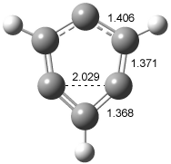
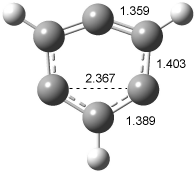
In order to confirm that this IR spectra comes from 2, Sander computed the structure and IR frequencies of both 1 and 2. The 2A1 structure of 1 had been studied previously2, but what had gone unnoticed is that another structure is possible, the 2B2 state. These two states differ in the separation between C1 and C3. When the distance is short, the SOMO is of a1 symmetry and this orbital has bonding character between these two carbon centers, giving rise to the 2A1 state (1a). As the distance gets longer between C1 and C3, a b2 orbital, having antibonding character between C1and C3, becomes lower in energy than the a1 orbital, so that the structure is 2B2 (1b). The UBLYP/cc-pVTZ optimized structures are shown in Figure 1. 1a is 2-3 kcal mol-1 lower in energy than 1b. Furthermore, 1b has one imaginary frequency and is not a local energy minimum. Sander also optimized the structures of 2a and 2b¸ finding little effect due to the fluorine substitution.

1a

1b

Figure 1. UBLYP/cc-pVTZ optimized structures of 1a (2A1) and 1b (2B1).

The computed IR frequencies are listed in Table 1. The computed frequencies (and their relative intensities) of 2a match up strikingly well with those of the experiment. Sander concludes that 2a has in fact been prepared and characterized.

References

(1) Venkataramani, S.; Winkler, M.; Sander, W., "Trifluoro-1,3,5-tridehydrobenzene," Angew. Chem. Int. Ed. 2007, 46, 4888-4893, DOI: 10.1002/anie.200700536

(2) Cristian, A. M. C.; Shao, Y.; Krylov, A. I., "Bonding Patterns in Benzene Triradicals from Structural, Spectroscopic, and Thermochemical Perspectives," J. Phys. Chem. A 2004, 108, 6581-6588, DOI: 10.1021/jp049007j.

InChI:

1: InChI=1/C6H3/c1-2-4-6-5-3-1/h1,4-5H
2: InChI=1/C6F3/c7-4-1-5(8)3-6(9)2-4

benzynes &DFT Steven Bachrach 20 Aug 2007 No Comments

Predicting the structure of artarborol

Here’s one more nice application of computationally-derived NMR chemical shifts towards solving a structure. Fattarusso and co-workers1 identified a component of wormwood called artarborol. COSY and ROESY experiments allowed for deducing four possible diasereomeric structures of artarborol, 1-4.

They then took two computational approaches towards resolving the structure. First, they performed an MM search for low energy conformers of 1-4. These conformers were then screened for those having a dihedral angle of around 90° for the C-8 and C-9 protons, due to a low couple constant for between these protons. Only conformers of 1 and 3 satisfied this criterion. An intense couple of the H-1 and H-5 protons indicated a transannular arrangement, and only conformers of 1 satisfy this criterion.

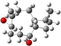
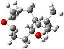
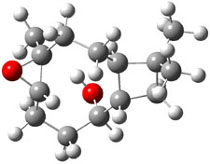
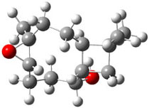
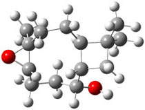
The second computational approach was to optimize some of the low energy conformers of 1 and 3 at mPW1PW91/6-31G(d,p) and compute their 13C chemical shifts. The five low energy conformers, two of 1 and three of 3, are shown in Figure 1. The resulting chemical shifts were averaged according to a Boltzmann distribution. These computed chemical shifts were then fit against the experimental values. The correlation factor for the computed shifts for 1 (r2=0.9997) was much better than that of 3 (r2=0.9713). The average deviation of the chemical shifts (after being corrected using the fitting procedure from the above correlation) was only 0.8ppm for 1 but 2ppm for 3. They therefore conclude that the structure of artarborol is 1.

1a
xyz

1b
xyz


3a
xyz

3b
xyz

3c
xyz

Figure 1. mPW1PW91 optimized conformations of possible artarborol diasteromers.1

References

(1) Fattorusso, C.; Stendardo, E.; Appendino, G.; Fattorusso, E.; Luciano, P.; Romano, A.; Taglialatela-Scafati, O., "Artarborol, a nor-Caryophyllane Sesquiterpene Alcohol from Artemisia arborescens. Stereostructure Assignment through Concurrence of NMR Data and Computational Analysis," Org. Lett., 2007, 9, 2377-2380, DOI: 10.1021/ol070803s.

(2) I thank Professor Ernesto Fattorusso for supplying me with the optimized coordinates of these compounds.

InChI

1: InChI=1/C14H24O2/c1-13(2)8-9-10(13)6-7-14(3)12(16-14)5-4-11(9)15/h9-12,15H,4-8H2,1-3H3/t9-,10-,11-,12-,14+/m0/s1

DFT &NMR Steven Bachrach 13 Aug 2007 No Comments

« Previous PageNext Page »