Archive for the 'Molecules' Category

Dihydroxycarbene

Following on the great study of hydroxycarbene1 (see my blog post), Schreiner now reports on the synthesis and characterization of dihydroxycarbene 1.2 It is prepared by high-vacuum flash pyrolysis of oxalic acid (Scheme 1).

Scheme 1

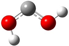
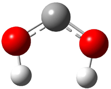
Dihydroxycarbene can exist in three different conformations characterized by the relationship about the C-O bond, either s-cis or s-trans. The three conformations are shown in Figure 1, and the s-trans,s-trans structure is the local energy minimum (computed at CCSD(T)/cc-pVTZ).

1tt (0.0)

1ct (0.1)

1cc (6.7)

Figure 1. CCSD(T)/cc-pVTZ optimized geometries and relative energies (kcal mol-1) of the conformers of 1.2

Identification of the 1 is made through comparison of the experimental and computed IR vibrational frequencies. As an example, the experimental and computed frequencies for the s-trans,s-trans conformer are listed in Table 1. The agreement is excellent.

Table 1. Computed and experimental vibrational frequencies (cm-1) and intensities (in parentheses) of the s-trans,s-trans conformation of 1.2


vibration

computed

experiment

1

3876.4 (23.5)

3633.2 / 3628.6 (w)

2

3871.4 (234.1)

3625.1 (s)

3

1443.1 (124.4)

1386.2 (m)

4

1370.5 (58.3)

1289.0 / 1287.4 (w)

5

1157.8 (470.6)

1110.3 / 1109.3 (vs)

6

1156.6 (1.4)


7

742.4 (178.8)

706.6 (s)

8

672.4 (0.0)


9

641.6 (11.2)


Unlike hydroxycarbene, dihydroxycarbene is stable. The amazing instability of hydroxycarbene is due to tunneling through a large barrier: nearly 30 kcal mol-1.1 The tunneling route for the decomposition of 1 is more difficult for two reasons. First, its C-O bond is quite strong; the C-O distance is quite short, 1.325 Å. This makes a long distance that must be traversed in the tunneling mode. (The strong bond is due to π-donation from the oxygen lone pair into the empty carbon p orbital; this is noted by the large rotational barrier about the C-O bonds of 17 kcal mol-1!) Second, the activation barrier for decomposition is very high, at least 34 kcal mol-1.

References

(1) Schreiner, P. R.; Reisenauer, H. P.; Pickard Iv, F. C.; Simmonett, A. C.; Allen, W. D.; Matyus, E.; Csaszar, A. G., "Capture of hydroxymethylene and its fast disappearance through tunnelling," Nature, 2008, 453, 906-909, DOI: 10.1038/nature07010.

(2) Schreiner, P. R.; Reisenauer, H. P., "Spectroscopic Identification of Dihydroxycarbene13," Angew. Chem. Int. Ed., 2008, 47, 7071-7074, DOI: 10.1002/anie.200802105

InChIs

1: InChI=1/CH2O2/c2-1-3/h2-3H

carbenes &Schreiner &Tunneling Steven Bachrach 06 Oct 2008 2 Comments

An update on Hydroxymethylene

A nice summary of the tunneling behavior of hydroxymethylene1 was just published by Bucher in Angewandte Chemie.2 Bucher strongly points out that the really novel part of this work is the very large barrier through which the proton tunnels. My blog post on this topic is here.

References

(1) Schreiner, P. R.; Reisenauer, H. P.; Pickard IV, F. C.; Simmonett, A. C.; Allen, W.
D.; Matyus, E.; Csaszar, A. G., "Capture of hydroxymethylene and its fast disappearance through tunnelling," Nature, 2008, 453, 906-909, DOI: 10.1038/nature07010.

(2) Bucher, G.; “Hydroxycarbene: Watching a Molecular Mole at Work,” Angew. Chem. Int. Ed., 2008, 47, 6957 – 6958, DOI: 10.1002/anie.200803195

carbenes &Schreiner &Tunneling Steven Bachrach 28 Aug 2008 No Comments

Cysteine conformers

Alonso and coworkers have developed the technique of laser ablation molecular beam Fourier transform microwave spectroscopy to detect biomolecules. In a recent paper1 they determined the structure of the glycine:one water complex – it is of the neutral configuration. They have now examined the conformations of cysteine2. The presence of the thiol side group adds considerable complexity to the problem due to the many conformations possible.

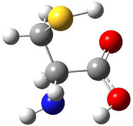
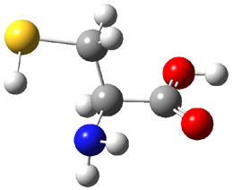
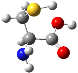
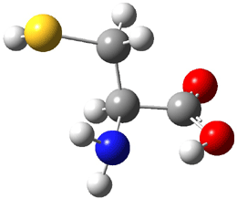
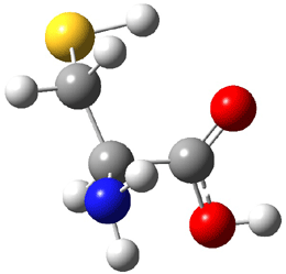
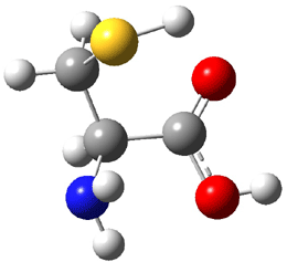
The experiment detected six conformers. Determining the structures responsible for each set of signals was made possible by comparing the experimental results with those determined by computation. Alonso computed 11 low energy conformations of cysteine at MP2/6-311++G(d,p). Then comparing the computed rotational constants and 14N nuclear quadrupole coupling tensor components with the experiment, they were able to match up all six experimental conformers with computed structures. The experimental and computed constants for the three most abundant structures are listed in Table 1. The geometries of all six conformers are drawn in Figure 1.

Table 1.Experimental and computed spectroscopic constants (MHz) for the three most abundant conformers of cysteine.2


IIb

Ia

Ib


Expt

MP2

Expt

MP2

Expt

MP2

A

3071.14

3040

4235.63

4221

2889.45

2855

B

1606.54

1623

1187.28

1185

1623.00

1664

C

1331.80

1347

1003.11

1013

1367.83

1386

χaa

-3.12

-3.14

-4.26

-4.67

-0.14

-0.01

χbb

2.44

2.59

2.78

2.86

0.44

0.25

χcc

0.68

0.55

1.49

1.80

-0.30

-0.24

ΔEa


0


450


325

aRelative energy in cm-1 computed at MP4/6-311++G(d,p)// MP2/6-311++G(d,p).

IIb (0.0)

Ia (450)

Ib (325)

IIa (527)

IIIβc (765)

IIIβb (585)

Table 1. Optimized structures of the six observed conformers of cysteine. Relative energies in cm-1 computed at MP4/6-311++G(d,p)//MP2/6-311++G(d,p). (Note – the geometries shown were optimized at PBE1PBE/6-311+G(d,p) since they MP2 structures are not available!)

This study demonstrates the nice complementary manner in which computation and experiment can work together in structure determination.

References


(1) Alonso, J. L.; Cocinero, E. J.; Lesarri, A.; Sanz, M. E.; López, J. C., "The Glycine-Water
Complex," Angew. Chem. Int. Ed. 2006, 45, 3471-3474, DOI: 10.1002/anie.200600342

(2) Sanz, M. E.; Blanco, S.; López, J. C.; Alonso, J. L., "Rotational Probes of Six Conformers of Neutral Cysteine," Angew. Chem. Int. Ed. 2008, DOI: 10.1002/anie.200801337

InChI

Cysteine: InChI=1/C3H7NO2S/c4-2(1-7)3(5)6/h2,7H,1,4H2,(H,5,6)/t2-/m0/s1
InChIKey: XUJNEKJLAYXESH-REOHCLBHBU

amino acids Steven Bachrach 26 Aug 2008 2 Comments

Hydroxymethylene tunnels through a large barrier

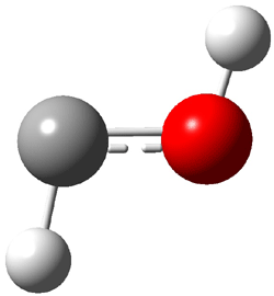
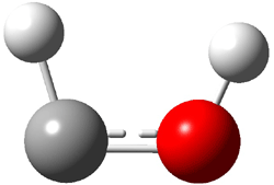
The very simple carbene hydroxymethylene, HOCH, has finally been prepared and characterized.1 Glyoxylic acid CHOCO2H is subjected to high-vacuum laser photolysis. It fragments into HOCH, which is then trapped into an argon matrix. The experimental IR frequencies match up very well with the CCSD(T)/cc-pVQZ harmonic frequencies of the trans isomer 1t that are also adjusted for anharmonic effects. The computed vertical excitation energy of 415 nm matches well with the experimental value of the maximum absorption in the UV/vis spectra of 427 nm.

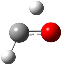
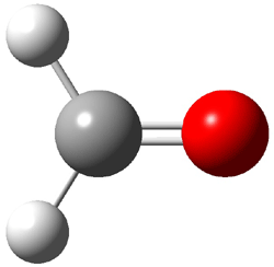
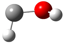
The other very interesting experimental result is that HOCH has a lifetime of about 2 hours in the matrix, while the deuterated species DOCH is stable. To explain these results, Schreiner, Allen and co-workers optimized a number of structures on the PES at CCSD(T)/cc-pVQZ and computed their energies using the focal point technique. The optimized structures and their relative energies are given in Figure 1.

1t (0.0)

TS2 (29.7)

2 (-52.1)

TS1(26.8)


1c (4.4)


Figure 1. Optimized CCSD(T)/cc-pVQZ structures of HOCH isomers and their Focal Point relative energies (kcal mol-1).1

The barriers for rearrangement from 1t are both very high. Rearrangement to formaldehyde 2 requires crossing a barrier of 29.7 kcal mol-1, while the barrier to convert to the cis isomer 1c is 26.8 kcal mol-1. (Note that from 1c a cleavage into CO and H2 can occur, but this barrier is another 47.0 kcal mol-1.) These barriers are too large to be crossed at the very low temperatures of the matrices. However, using the intrinsic reaction potential at CCSD(T)/cc-pVQZ and WKB theory, the tunneling lifetime of HOCH is computed to be 122 minutes, in excellent accord with the experiment. The lifetime for DOCH is computed to be over 1200 years. Thus, the degradation of hydroxymethylene is entirely due to tunneling through a very large classical barrier! This rapid tunneling casts serious doubt on the ability to ever identify any hydroxymethylene in interstellar space.

References

(1) Schreiner, P. R.; Reisenauer, H. P.; Pickard IV, F. C.; Simmonett, A. C.; Allen, W.
D.; Matyus, E.; Csaszar, A. G., "Capture of hydroxymethylene and its fast disappearance through tunnelling," Nature, 2008, 453, 906-909, DOI: 10.1038/nature07010.

InChI

1: InChI=1/CH2O/c1-2/h1-2H
2: InChI=1/CH2O/c1-2/h1H2

carbenes &focal point &Schreiner &Tunneling Steven Bachrach 19 Aug 2008 4 Comments

Dihydrodiazatetracene: is it antiaromatic?

Schleyer continues his study of aromaticity with a paper1 that picks up on the theme presented in one2 I have previously blogged on – the relationship between a formally aromatic pyrazine and formally antiaromatic dihydropyrazine. He now examines the diazotetracene 1 and it dihydro analogue 2.1 In terms of formal electron count, 1 should be aromatic, just like the all carbon analogue tetracene 3, and 2 should be antiaromatic.

Schleyer used the NICSπzz values obtained in the center of each ring to evaluate the aromatic/antiaromatic character of these three molecules. These calculations were performed using canonical molecular orbitals and repeated using localized molecular orbitals. The results are similar for each method, and the canonical MO values are presented in Table 1. As expected for an aromatic compound, each ring of tetracene 3 has large negative NICS values, indicating that each ring is locally aromatic and the molecule as a whole is aromatic. The same is true for the diazotetracene 1. (In fact the NICS values for 1 and 3 are remarkably similar.) However, for 2, the dihydropyrazine ring has a positive NICS values, indicative of a locally antiaromatic ring. While the three phenyl rings have negative NICS values, these absolute values are smaller than for the rings of 1 or 3, indicating an attenuation of their aromaticity. Nonetheless, the sum of the NICS values of 2 is negative, suggesting that the molecule is globally aromatic, though only marginally so. This is due to the antiaromaticity of the dihydropyrazine ring being delocalized to some extent over the entire molecule. Schleyer, concludes that “large 4n π compounds […] are not appreciably destabilized relative to their 4n+2 π congeners.”

Table 1 NICSπzz (ppm) for each ring of 1-3 and their sum.1


1

-30.0

-42.5

-41.1

-30.1

sum = -144.0


2

-26.3

-14.2

31.3

-16.7

sum = -25.9


3

-29.6

-42.1

-42.1

-29.6

Sum = -143.4

References

(1) Miao, S.; Brombosz, S. M.; Schleyer, P. v. R.; Wu, J. I.; Barlow, S.; Marder, S. R.; Hardcastle, K. I.; Bunz, U. H. F., "Are N,N-Dihydrodiazatetracene Derivatives Antiaromatic?," J. Am. Chem. Soc., 2008, 130, 7339-7344, DOI: 10.1021/ja077614p.

(2) Miao, S.; Schleyer, P. v. R.; Wu, J. I.; Hardcastle, K. I.; Bunz, U. H. F., "A Thiadiazole-Fused N,N-Dihydroquinoxaline: Antiaromatic but Isolable," Org. Lett. 2007, 9, 1073-1076, DOI: 10.1021/ol070013i

InChIs

1: InChI=1/C18H12/c1-2-6-14-10-18-12-16-8-4-3-7-15(16)11-17(18)9-13(14)5-1/h1-12H

2: InChI=1/C16H10N2/c1-2-6-12-10-16-15(9-11(12)5-1)17-13-7-3-4-8-14(13)18-16/h1-10H

3: InChI=1/C16H12N2/c1-2-6-12-10-16-15(9-11(12)5-1)17-13-7-3-4-8-14(13)18-16/h1-10,17-18H

Aromaticity &polycyclic aromatics &Schleyer Steven Bachrach 15 Jul 2008 No Comments

Hexacoordinate carbon

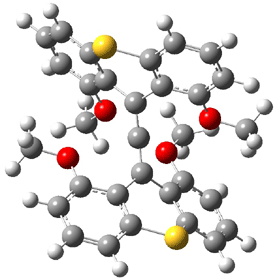
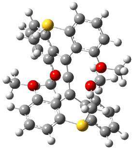
The search for the elusive hypervalent carbon atom took an interesting turn for the positive with the report of the synthesis and characterization of 1 and especially its dication 2.1 The x-ray structure was obtained for both compounds along with computing their B3PW91/6-31G(d) geometries. These computed geometries are shown in Figure 1.


1


2

1

2

Figure 1. B3PW91/6-31G(d) optimized geometries of 1 and 2.1

The allene fragment is bent in both structures: 168.5° (169.9° at B3PW91) in 1 and 166.8° (172.7° at B3PW91) in 2. The distances between the central carbon atom of the allene and the four oxygen atoms are 2.66 to 2.82 Å, and computed to be a little longer. Interestingly, these distance contract when the dication 2 is created; ranging from 2.64 to 2.75 Å (again computed to be a little longer). These distances, while significantly longer than normal covalent C-O bonds, are less than the sum of the C and O van der Waals radii. But are they really bonds?

This is not a trivial answer to solve. The authors opt to employ topological electron density analysis (Bader’s atoms-in-molecules approach). Using the electron density from both the high resolution x-ray density map and from the DFT computations, bond paths between the central allene carbon and each oxygen are found, though with, as expected, low values of ρ. The Laplacian of the density at the critical point are positive, indicative of ionic interactions. So according to Bader’s model, the existence of a bond path in a ground state molecule is the necessary and sufficient condition for bonding.

The others conclude by proposing that OC intermolecular interactions with separations of around 2.6 to 2.8 Å may also suggest hypervalent cases. They note that about 2000 structures in the Cambridge crystallographic database fit this criterion.

References

(1) Yamaguchi, T.; Yamamoto, Y.; Kinoshita, D.; Akiba, K.-y.; Zhang, Y.; Reed, C. A.; Hashizume, D.; Iwasaki, F., "Synthesis and Structure of a Hexacoordinate
Carbon Compound," J. Am. Chem. Soc., 2008, 130, 6894-6895, DOI: 10.1021/ja710423d.

InChIs

1: InChI=1/C31H24O4S2/c1-32-20-9-5-13-24-28(20)18(29-21(33-2)10-6-14-25(29)36-24)17-19-30-22(34-3)11-7-15-26(30)37-27-16-8-12-23(35-4)31(19)27/h5-16H,1-4H3
InChiKey= RJBVLFYVIBCWKW-UHFFFAOYAD

2: InChI=1/C33H30O4S2/c1-34-22-11-7-15-26-30(22)20(31-23(35-2)12-8-16-27(31)38(26)5)19-21-32-24(36-3)13-9-17-28(32)39(6)29-18-10-14-25(37-4)33(21)29/h7-18H,1-6H3/q+2
InChIKey= MVHOSIVPHPDYJJ-UHFFFAOYAM

DFT &non-classical Steven Bachrach 03 Jun 2008 3 Comments

Protonated acetylene

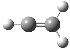
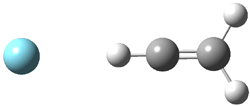
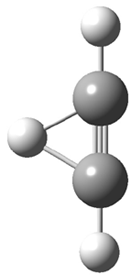
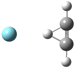
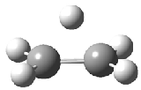
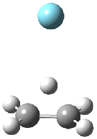
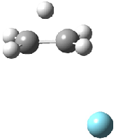
Duncan and Schleyer1 have investigated protonated acetylene and the protonated acetylene dimer. These ions are created in a pulsed supersonic nozzle/pulsed electrical discharge with a weakly bound argon atom as a tag. IR laser photodissociation spectroscopy allows for the detection of peaks down to 2000 cm-1, a region not previously explored for this cation. The experimental IR spectrum for H+(C2H2).Ar has two main features: at 3146 and 2217 cm-1. The 3146 cm-1 corresponds to the previously observed peak2 at 3142 cm-1 and is similar to the absorption in acetylene (3136 cm-1). MP2/6-311+G(2d,2p) computations were performed on the classical and non-classical structures of H+(C2H2), with and without a complexed argon atom. These geometries are displayed in Figure 1 and the predicted vibrational frequencies are listed in Table 1.

Figure 1. MP2/6-311+

Table 1. Relative energies (kcal mol-1) and frequencies of protonated acetylene
and the protonated acetylene-argon cluster.1


Rel E

Frequencies (scaled)

H+(acetylene)Ar non-classical

0.0

3139, 2123

H+(acetylene)Ar classical

7.8

3084, 2954, 2878, 1673

H+(acetylene) non-classical

0.0

3219, 2250

H+(acetylene) classical

7.1

3162, 29947, 2874

Experiment


3364, 3212, 3146, 2217


The argon tag only slightly perturbs the spectrum, as expected for a weakly bond atom remote from most of the hydrogen atoms. The predicted spectra of the two non-classical ions are in nice agreement with the experiment – particularly the interesting peak at 2123 cm-1 that is due to the bridged proton. This spectra, and the confirmation of the bridging, non-classical structure, makes a nice pair with the recently reported bridging, non-classical structure of the ethyl cation,3 which I blogged on previously.

The spectrum of the H+(C4H4) ion show a doublet at 3129 and 3158 cm-1 and two small peaks at 1261 and 1365 cm-1. The computed structure that comes closest to matching this spectrum is for the asymmetrically bridged dimer (See Figure 2), though is much more energetic than its isomers. The authors speculate that the bridged dimer is trapped in an energy-well during the thermal expansion, which prevents the formation of the lower energy isomers.

Figure 2. Schematic drawing and relative energies of the H+(C4H4) ion.
(Note – unfortunately the authors have supplied insufficient information in the Supporting Materials to completely define the geometries of these molecules!)

References

(1) Douberly, G. E.; Ricks, A. M.; Ticknor, B. W.; McKee, W.
C.; Schleyer, P. v. R.; Duncan, M. A., "Infrared Photodissociation
Spectroscopy of Protonated Acetylene and Its Clusters," J. Phys. Chem. A, 2008, 112, 1897-1906, DOI: 10.1021/jp710808e.

(2) Gabrys, C. M.; Uy, D.; Jagod, M. F.; Oka, T.; Amano, T., "Infrared Spectroscopy of Carboions. 8. Hollow Cathode Spectroscopy of Protonated Acetylene, C2H3+," J. Phys. Chem., 1995, 99, 15611-15623, DOI: 10.1021/j100042a042.

(3) Andrei, H.-S.; Solcà, N.; Dopfer, O., "IR Spectrum of the Ethyl Cation: Evidence
for the Nonclassical Structure," Angew. Chem. Int. Ed., 2008, 47, 395-397, DOI: 10.1002/anie.200704163

ethyl cation &Schleyer Steven Bachrach 01 May 2008 No Comments

m-Xylylene

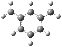
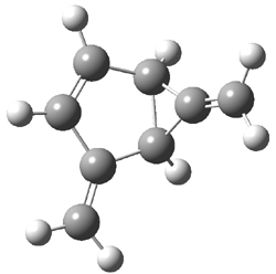
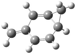
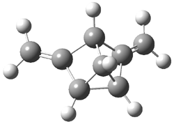
The photochemistry of m-xylylene 2 has been studied by Sander1 and, as might be anticipated, it’s fascinating! Flash vapor pyrolysis of 1 produces 2. Photolysis of 2 at wavelengths above 400nm gives 3 and 4, while photolysis at 254 nm gives 5. These are products are novel strained hydrocarbons. Confirmation of their structures was obtained by comparing their experimental IR spectra with that computed at B3LYP/6-311G(d,p). Table 1 compares the experimental and computed IR absorptions for 2-5. Note in particular the fine agreement between the two, especially the predicted changes due to i>d4 substitution for all the phenyl positions.

Table 1. Experimental and Calculated vibrational frequencies (cm-1) of 2-5.1


Mode

ν (expt)

ν (calc)

ν (expt)

ν (calc)


2

2-d4

11

640.5

655.0

645.6

661.3

12

723.9

721.0

581.0

576.5

15

766.4

777.0

759.1

772.6

16

834.9

849.0

831.4

847.3


3

3-­d4

11

733.6

757.8

681.1

698.8

16

869.9

899.9

808.1

822.2

17

883.2

915.3

703.4

726.5

33

1640.6

1696.4

1614.4

1658.8


4

4-­d4

10

706.6

715.8

627.1

633.9

11

757.4

770.8

689.7

702.3

15

874.2

896.6

763.5

784.2

23

1065.2

1082.7

1001.4

1017.5


5

5-­d4

10

742.9

764.2

660.5

676.4

16

851.7

876.4

787.2

797.5

17

852.9

880.1

789.8

799.9

33

1678.6

1747.1

166.2

1709.7

34

1683.8

1758.7

1669.2

1719.9


The computed structures of 2-5 and their relative energies are shown in Figure 1. Triplet 1 is the lowest energy isomer, with the singlet-triplet gap of 6.22 kcal mol-1. This compares with recent high-level computations which give a value of 13.8 kcal mol-1. 2 The other structures are much higher in energy. These other isomers have unusual bonding environments – 3 contains the strained methylenecyclopropane group, 4 is an anti-Bredt compound, and 5 is a very strained tricycle. These compounds can only be prepared by the application of light to provide the energy needed for their creation.

2
Triplet 0.0
Singlet 6.22

3
19.20

4
25.74

5
48.64

Figure 1. B3LYP/6-311G(d,p) optimized structures of 2-5 and their relative energies (kcal mol-1).1

References

(1) Neuhaus, P.; Grote, D.; Sander, W., "Matrix Isolation, Spectroscopic Characterization, and Photoisomerization of m-Xylylene," J. Am. Chem. Soc., 2008, 130, 2993-3000, DOI: 10.1021/ja073453d.

(2) Wang, T.; Krylov, A. I., "The effect of substituents on electronic states’ ordering in meta-xylylene diradicals: Qualitative insights from quantitative studies," J. Chem. Phys., 2005, 123, 104304, DOI: 10.1063/1.2018645.

InChIs

2: InChI=1/C8H8/c1-7-4-3-5-8(2)6-7/h3-6H

3: InChI=1/C8H8/c1-5-3-4-7-6(2)8(5)7/h3-4,7-8H,1-2H2; InChIKey=IAJQBWKVTDKBHW-UHFFFAOYAA

4: InChI=1/C8H8/c1-6-2-3-7-5-8(7)4-6/h2-4,7H,1,5H2; InChIKey=XSFFUTQMPAAWHN-UHFFFAOYAU

5: InChI=1/C8H8/c1-3-5-4(2)7-6(3)8(5)7/h5-8H,1-2H2; InChIKey=USZJYFGTTGREFL-UHFFFAOYAB

DFT &diradicals Steven Bachrach 09 Apr 2008 1 Comment

Ethyl cation

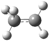
The structure of the simple, fundamental ethyl cation has finally been ascertained. Computational studies had long suggested the non-classical structure 1 for this cation. The classical structure 2 is a transition state for scrambling the protons. The MP2/6-311G(2d,p) geometries of both structures are shown in Figure 1.

1

2

1.Ar(C2v)

1.Ar(Cs)

Figure 1. MP2/6-311G(2d,f) structures of 1, 2, 1.Ar(C2v) and 1.Ar(Cs).

Dopfer1 has now obtained IR spectrum of ethyl cation by single-photon IR photodissociation spectroscopy through the reaction

C2H5+ . Ar + hν → C2H5+ + Ar

Two structures of the ethyl cation associated with Ar were optimized at MP2/6-311G(2df,2pd). (The MP2/6-311G(2d,p) structures are shown in Figure 1.) Both of their computed IR spectra have stretches at nearly identical wavenumbers as for ethyl cation 1 itself. The experimental IR spectra has absorptions at 3317 and 3037 cm-1, very close to the computed frequencies for 1.Ar(C2v). This provides strong experimental evidence that ethyl cation is in fact a non-classical ion.

References

(1) Andrei, H.-S.; Solcà, N.; Dopfer, O., "IR Spectrum of the Ethyl Cation: Evidence for the Nonclassical Structure," Angew. Chem. Int. Ed. 2008, 47, 395-397, DOI: 10.1002/anie.200704163

ethyl cation Steven Bachrach 27 Feb 2008 3 Comments

Oxime Bond Dissociation Energies

The bond dissociation energies (BDE) of the O-H bond of oximes R1R2C=N-OH) are discussed in Chapter 2.1.1.2. The controversy associated with these values originates from conflicting experimental data coming from calorimetric and electrochemical experiments. Some of the conflicting data are listed in Table 1. The electrochemical method provides energies at least a couple of kcal mol-1 too large, sometimes much more than that. I described in the book some composite method computations (G3MP, G3, CBS-QB3 and CBS-ANO)1 that suggest the BDE of acetone oxime is around 85 kcal mol-1, consistent with the calorimetric results. These authors could not apply these expensive methods to other compounds and were forced to use UB3LYP, which underestimates the values of the BDEs.

Table 1. BDEs (kcal mol-1) from calorimetric and electrochemical experiments
and ONIOM-G3B3 computations.


R1

R2

Calorimetric

Electrochemical

ONIOM-G3B3

Me

Me

84.3a

95.8b

85.7

Ph

Ph

82.4c

89.0b

81.8

i-Pr

i-Pr

79.7c

87.7d

83.0

t-Bu

i-Pr

82.6c

86.0e

83.0

t-Bu

t-Bu

79.2c

84.2,d
82.6e

78.7

t-Bu

1-Ad

79.2c

81.7d

78.7

fluorenyl

82.0c

87.5b

80.4


aRef. 2. bRef. 3. cRef. 1. dRef. 4. eRef. 5

Fu6 has now applied the ONIOM-G3B37 approach to this problem. This is a clever way of attacking large molecules that require rather large computations to appropriately treat the quantum mechanics. So, each step of the G3B3 composite method is split into two levels: the high level is computed with the appropriate method from the G3B3 procedure, while the low level is treated with B3LYP. These resulting BDEs are listed in Table 1 and show remarkably nice agreement with the calorimetric results. These computed BDEs confirm that the electrochemical results are in error. Fu also computed the BDEs of some 30 other oximes for which electrochemical BDEs are available and for the large majority of these compounds, the electrochemical values are again too large.

References

(1) Pratt, D. A.; Blake, J. A.; Mulder, P.; Walton, J. C.; Korth, H.-G.; Ingold, K. U., "O-H Bond Dissociation Enthalpies in Oximes: Order Restored," J. Am. Chem. Soc. 2004, 126, 10667-10675, DOI: 10.1021/ja047566y.

(2) Mahoney, L. R.; Mendenhall, G. D.; Ingold, K. U., "Calorimetric and Equilibrium Studies on Some Stable Nitroxide and Iminoxy Radicals. Approximate Oxygen-Hydrogen Bond Dissociation Energies in Hydroxylamines and Oximes," J. Am. Chem. Soc. 1973, 95, 8610-8614, DOI: 10.1021/ja00807a018.

(3) Bordwell, F. G.; Ji, G.-Z., "Equilibrium Acidities and Homolytic Bond Dissociation Energies of the H-O Bonds in Oximes and Amidoximes," J. Org. Chem. 1992, 57, 3019 – 3025, DOI: 10.1021/jo00037a014.

(4) Bordwell, F. G.; Zhang, S., "Structural Effects on Stabilities of Iminoxy Radicals," J. Am. Chem. Soc. 1995, 117, 4858-4861, DOI: 10.1021/ja00122a016.

(5) Bordwell, F. G.; Liu, W.-Z., "Solvent Effects on Homolytic Bond Dissociation Energies of Hydroxylic Acids," J. Am. Chem. Soc. 1996, 118, 10819-10823, DOI: 10.1021/ja961469q.

(6) Chong, S. S.; Fu, Y.; Liu, L.; Guo, Q. X., "O-H Bond Dissociation Enthalpies of Oximes: A Theoretical Assessment and Experimental Implications," J. Phys. Chem. A 2007, 111, 13112-13125, DOI: 10.1021/jp075699a.

(7) Li, M. J.; Liu, L.; Fu, Y.; Guo, Q. X., "Development of an ONIOM-G3B3 Method to Accurately Predict C-H and N-H Bond Dissociation Enthalpies of Ribonucleosides and Deoxyribonucleosides," J. Phys. Chem. B 2005, 109, 13818-13826, DOI: 10.1021/jp0508204.

Bond Dissociation Energy &oximes Steven Bachrach 22 Jan 2008 No Comments

« Previous PageNext Page »