Tunneling within a phenylnitrene

Tunneling Steven Bachrach 15 Dec 2016 No Comments

Reva and McMahon report a very nice experimental and computational study implicating hydrogen atom tunneling in the rearrangement of the nitrene 1 into the ketene 2.1 The reaction is carried out by placing azide 3 in an argon matrix and photolyzing it. The IR shows that at first a new compound A is formed and that over time the absorptions of A erode and those of a second compound B grow in. This occurs whether the photolysis continues or not over time.

IR spectra were computed at B3LYP/6-311++G(d,p) for compounds 31 and 2 and they match up very well with the recorded spectra of A and B, respectively. The triplet state of nitrenes are typically about 20 kcal mol-1 lower in energy than the singlet states. The EPR spectrum confirms that 1 is a triplet.

So how does the conversion of 31 into 2 take place, especially at 10 K? The rate constant for this conversion at 10 K is estimated as 1 x 10⁻⁵ s-1, which implies a barrier from classical transition state theory of only 0.2 kcal mol-1. That low a barrier seems preposterous, and suggests that the reaction may proceed via tunneling. This notion is supported by the experiment on the deuterated analogue, which shows no conversion of 1D into 2D.

The authors propose that 31 undergoes a hydrogen migration on the triplet surface through transition state 34 to give 32, which then undergoes intersystem crossing to give singlet 2. The structures of these critical points calculated at B3LYP/6-311++G(d,p) are shown in Figure 1. The computed activation barrier is 20.7 kcal mol-1. (The barrier height ranges from 16.7 to 23.0 with a variety of different computational methods.) This large barrier precludes a classical over-the-top reaction and points towards tunneling. The barrier width is estimated at about 2.1 Å. WKB computations estimate the tunneling half time of about 21 min, somewhat smaller than in the experiments, and the estimate for the deuterated species is 150,000 years.

31

34

32

Figure 1. B3LYP/6-311++G(d,p) optimized structures of 31, 32, and the TS 34.

References

1) Nunes, C. M.; Knezz, S. N.; Reva, I.; Fausto, R.; McMahon, R. J., "Evidence of a Nitrene Tunneling Reaction: Spontaneous Rearrangement of 2-Formyl Phenylnitrene to an Imino Ketene in Low-Temperature Matrixes." J. Am. Chem. Soc. 2016, 138, 15287-15290, DOI: 10.1021/jacs.6b07368.

InChIs:

1: InChI=1S/C7H5NO/c8-7-4-2-1-3-6(7)5-9/h1-5H
InChIKey=QZTZBORTPUZAGF-UHFFFAOYSA-N

2: InChI=1S/C7H5NO/c8-7-4-2-1-3-6(7)5-9/h1-4,8H
InChIKey=ZWHBMBVIYUVTGT-UHFFFAOYSA-N

NMR coupling constants of strychnine

NMR Steven Bachrach 15 Nov 2016 No Comments

Helgaker, Jaszunski, and Swider1 have examined the use of B3LYP with four different basis sets to compute the spin-spin coupling constants in strychnine 1.


1

They used previously optimized coordinates of the two major conformations of strychnine, shown in Figure 1.

Conformer A

Conformer B

Figure 1. Confrmations of strychnine 1.

They tested four basis sets designed for NMR computations: pcJ-0,2 pcJ-1,2 6-31G-J,3 and 6-311G-J.3 pCJ-0 and 6-31G-J are relatively small basis sets, while the other two are considerably larger.

All four basis sets provide values of the 122 J(C-H) with a root mean square deviation of less than 0.6 Hz. J(HH) and J(CC) coupling constants are also well predicted, especially with the larger pcJ-1 basis set. They also examined the four Ramsey terms in the coupling model. The Fermi contact term dominates, and if the large pcJ-1 basis set is used to calculate it, and the smaller pcJ-0 basis set is used for the other three terms, the RMS error only increases from 0.18 to 0.20 Hz. Taking this to the extreme, they omitted calculating any of the non-Fermi contact terms, with again only small increases in the RMS – even with the small pcJ-0 basis set. Considering the computational costs, one should seriously consider whether the non-Fermi contact terms and a small basis set might be satisfactory for your own problem(s) at hand.

References

1) Helgaker, T.; Jaszuński, M.; Świder, P., "Calculation of NMR Spin–Spin Coupling Constants in Strychnine." J. Org. Chem. 2016, ASAP, DOI: 10.1021/acs.joc.6b02157.

2) Jensen, F., "The Basis Set Convergence of Spin−Spin Coupling Constants Calculated by Density Functional Methods." J. Chem. Theor. Comput. 2006, 2, 1360-1369, DOI: 10.1021/ct600166u.

3) Kjær, H.; Sauer, S. P. A., "Pople Style Basis Sets for the Calculation of NMR Spin–Spin Coupling Constants: the 6-31G-J and 6-311G-J Basis Sets." J. Chem. Theor. Comput. 2011, 7, 4070-4076, DOI: 10.1021/ct200546q.

InChIs

Strychnine 1: InChI=1S/C21H22N2O2/c24-18-10-16-19-13-9-17-21(6-7-22(17)11-12(13)5-8-25-16)14-3-1-2-4-15(14)23(18)20(19)21/h1-5,13,16-17,19-20H,6-11H2/t13-,16-,17-,19-,20-,21+/m0/s1
InChIKey=QMGVPVSNSZLJIA-FVWCLLPLSA-N

More examples of structure determination with computed NMR chemical shifts

NMR &terpenes Steven Bachrach 25 Oct 2016 No Comments

Use of computed NMR chemical shifts in structure determination is really growing fast. Presented here are a couple of recent examples.

Nguyen and Tantillo used computed chemical shifts with the DP4 analysis to identify the structure of three terpenes 1-3.1 They optimized the geometries of all of the diastereomers of each compound, along with multiple conformations of each diastereomer, at B3LYP/6-31+G(d,p) and then computed the chemical shifts at SMD(CHCl3)–mPW1PW91/6-311+G(2d,p). The chemical shifts were Boltzmann weighted including all conformations within 3 kcal mol-1 of the lowest energy structure.

For 1, the DP4 analysis using just the proton shifts predicted a different isomer than using the carbon shifts, but when combined, DP4 predicted the structure, with 98.8% confidence, shown in the scheme above, and in Figure 1. For 2, the combined proton and carbon shift analysis with DP4 indicated a 100% confidence of the structure shown in the scheme and Figure 1. Lastly, for 3, which is more complicated due to the conformations of the 9-member ring, DP4 predicts with 100% confidence the structure shown in the scheme and Figure 1.

1

2

3

Figure 1. Optimized geometries of 1-3.

Feng, Davis and coworkers have examined a series of anthroquionones from Australian marine sponges.2 The structure of one compound was a choice of two options: 4 or 5. Initial geometries were obtain by molecular mechanics and the low energy isomers were then reoptimized at B3LYP/6-31+G(d,p). The chemical shifts were computed using PCM/MPW1PW91/6-311+G(2d,p). Application of the DP4 method indicate the structure to be 4 with a 100% confidence level. The lowest energy conformer of 4 is shown in Figure 2.

Figure 2. Optimized geometry of 4.

References

1) Nguyen, Q. N. N.; Tantillo, D. J. “Using quantum chemical computations of NMR chemical shifts to assign relative configurations of terpenes from an engineered Streptomyces host,” J. Antibiotics 2016, 69, 534–540, DOI: 10.1038/ja.2016.51.

2) Khokhar, S.; Pierens, G. K.; Hooper, J. N. A.; Ekins, M. G.; Feng, Y.; Rohan A. Davis, R. A. “Rhodocomatulin-Type Anthraquinones from the Australian Marine Invertebrates Clathria hirsuta and Comatula rotalaria,” J. Nat. Prod., 2016, 79, 946–953, DOI: 10.1021/acs.jnatprod.5b01029.

InChIs

1: InChI=1S/C15H24/c1-10-5-6-15(4)8-11-7-14(2,3)9-12(11)13(10)15/h9-11,13H,5-8H2,1-4H3/t10-,11+,13-,15+/m1/s1
InChIKey=KVSCZIPUFBVHBM-OICBVUGWSA-N

2: InChI=1S/C15H24/c1-10-5-6-15(4)8-11-7-14(2,3)9-12(11)13(10)15/h5,11-13H,6-9H2,1-4H3/t11-,12-,13+,15-/m0/s1
InChIKey=ZLYGJLHCPYVGDA-XPCVCDNBSA-N

3: InChI=1S/C20H32/c1-14-6-9-18-19(3,4)10-11-20(18,5)13-17-15(2)7-8-16(17)12-14/h6,13,15-16,18H,7-12H2,1-5H3/b14-6-,17-13-/t15-,16-,18-,20+/m0/s1
InChIKey=JZGOFJIAHJJJDK-ICZJPRMTSA-N

4: InChI=1S/C18H14O7/c1-7(19)13-10(20)6-11(21)15-16(13)17(22)9-4-8(24-2)5-12(25-3)14(9)18(15)23/h4-6,20-21H,1-3H3
InChIKey=MPQMZEXRJVMYBT-UHFFFAOYSA-N

5: InChI=1S/C18H14O7/c1-7(19)13-10(20)6-11(21)15-16(13)14-9(17(22)18(15)23)4-8(24-2)5-12(14)25-3/h4-6,20-21H,1-3H3
InChIKey=WIKIUXNPFURKNF-UHFFFAOYSA-N

Further development of DP4 for NMR structure determination

NMR Steven Bachrach 11 Oct 2016 No Comments

Computational chemistry has had a remarkable impact on the field of structure determination by NMR spectroscopy. The ability to efficiently compute 13C and 1H chemical shifts allows for comparison of the computed chemical shifts of potential structures against the experimental values, a tremendous aid in structure determination (see some examples in previous posts). Goodman and Smith developed the DP4 method1 (see this post) to assist in identifying proper structures by means of statistical distribution of errors and Bayes Theorem.

The Goodman group now reports on workflow solutions to structure prediction using DP4.2 They explore the use of open source computational tools both for predicting conformations and for computing the chemical shifts. They use a set of 10 drugs to test the performance. In general, the original DP4 method works very well in predicting drug structure, despite the fact that DP4 parameters were developed for natural products. The only failure is for simvastatin, where the large number of diastereomers and conformational flexibility prove to be too complex. The open source tools perform just slightly less effectively than the commercial packages, but are certainly a viable route for those with limited resources. The authors also provide a series of python scripts that allow users to create a seamless workflow; these should prove most helpful to the structure determination community.


Simvastatin

References

1) Smith, S. G.; Goodman, J. M. "Assigning Stereochemistry to Single Diastereoisomers by GIAO
NMR Calculation: The DP4 Probability," J. Am. Chem. Soc. 2010, 132, 12946-12959, DOI: 10.1021/ja105035r.

2) Ermanis, K.; Parkes, K. E. B.; Agback, T.; Goodman, J. M. “Expanding DP4: application to drug compounds and automation,” Org. Biomol. Chem., 2016, 14, 3943-3949, DOI: 10.1039/c6ob00015k.

InChIs

Simvastatin: InChI=1S/C25H38O5/c1-6-25(4,5)24(28)30-21-12-15(2)11-17-8-7-16(3)20(23(17)21)10-9-19-13-18(26)14-22(27)29-19/h7-8,11,15-16,18-21,23,26H,6,9-10,12-14H2,1-5H3/t15-,16-,18+,19+,20-,21-,23-/m0/s1
InChIKey=RYMZZMVNJRMUDD-HGQWONQESA-N

Polytriangulane

Schreiner Steven Bachrach 27 Sep 2016 1 Comment

Cyclopropyl rings can be joined together in a spiro fashion to form triangulanes. An interesting topology can be made by joining the rings to form a helical pattern, as shown in the [9]triangulane 1 below. Allen, Quanz, and Schreiner1 have examined the notion of an infinite helical molecule formed in this way.


1

First, they describe how one can generate the coordinates of such a beast using a closed analytical expression, which is a really nice demonstration of applied geometry. Next, they compute the geometry of a series of [n]triangulanes at M06-2x/6-31G(d). The geometries of [9]triangulane and their largest example, [42]triangulane 2 are shown in Figure 1.

1

2

Figure 1. M06-2x/6-31G(d) optimized geometries of 1 and 2.

They show that the geometry of 2 exhibits a structure that has two different C-C distances: one between the spiro carbons, and the second between the spiro carbon and the methylene carbon. The distance between the spiro carbons is rather short (1.458 Å), suggesting that the bonding here is between carbons that are nearly sp2-hybridized.

Lastly, they discuss the thermodynamics of polytriangulane. They employ a series of homodesmotic reactions to attempt to determine the enthalpy for adding another cyclopropyl ring to an extended triangulane. Unfortunately, the computed enthalpy is quite dependent on functional used. Similar attempts to define the strain energy is also flawed in this way. However, regardless of the functional the enthalpy for adding a cyclopropane ring appears to reach an asymptote rather quickly. So, using [3]triangulane they estimate that the strain energy per mole of cyclopropane in triangulane is about 42.7 kcal mol-1, or about 14 kcal mol-1 of strain due to the spiroannulation.

References

(1) Allen, W. D.; Quanz, H.; Schreiner, P. R. “Polytriangulane,” J. Chem. Theory Comput. 2016, 12, 4707–4716, DOI: 10.1021/acs.jctc.6b00669.

InChIs

1: InChI=1S/C19H22/c1-2-12(1)5-14(12)7-16(14)9-18(16)11-19(18)10-17(19)8-15(17)6-13(15)3-4-13/h1-11H2/t14-,15-,16-,17-,18-,19-/m0/s1
InChIKey=XBTZCZDSKVTALB-DYKIIFRCSA-N

Bergman Cyclization on a Gold Surface

Bergman cyclization Steven Bachrach 19 Sep 2016 No Comments

The Bergman cyclization and some competitive reactions are discussed in detail in Chapter 4 of by book. The Bergman cyclization makes the C1-C6 bond from an enediyne. Another, but rarer, option is to make the C1-C5 bond, the Schreiner-Pascal cyclization pathway. de Oteyza and coworkers have examined the competition between these two pathways for 1 on a gold surface, and used STM and computations to identify the reaction pathway.1

The two pathways are shown below. The STM images identify 1 as the reactant on the gold surface and the product is 6. No other product is observed.

Projector augmented wave (PAW) pseudo-potential computations using the PBE functional were performed for the reaction on a Au (111) surface was modeled by a 7 x 7 x 3 supercell. The optimized geometries of the critical points are show in Figure 1.

1

TS(1→2)

TS(1→3)

2

3

TS(2→6)

TS(3→5)

6

5

Figure 1. Optimized geometries of the critical points on the two reaction pathways.

Explicit values of the relative energies are not given in either the paper or the supporting information, but rather a plot shows the relative positions of the critical points. The important points are the following: (a) the barrier for the C1-C5 cyclization is lower than the barrier for the C1-C6 cyclization and 3 is lower in energy than 2; (b) 5 is lower in energy than 6; and (c) the barrier for taking 2 to 6 is significantly below the barrier taking 3 into 5. The barrier for the phenyl migration taking 3 into 5 is so high because of a strong interaction between the carbon radical and a gold atom of the surface. The authors suggest that the two initial cyclizations are reversible, but the very high barrier for forming 5 precludes it from taking place, leaving only the route to 6 as a viable pathway.

References

(1) de Oteyza, D. G.; Paz, A. P.; Chen, Y.-C.; Pedramrazi, Z.; Riss, A.; Wickenburg, S.; Tsai, H.-Z.; Fischer, F. R.; Crommei, M. F.; Rubio, A. “Enediyne Cyclization on Au(111),” J. Amer. Chem. Soc. 2016, 138, 10963–10967, DOI: 10.1021/jacs.6b05203.

InChIs

1: InChI=1S/C22H14/c1-3-9-19(10-4-1)15-17-21-13-7-8-14-22(21)18-16-20-11-5-2-6-12-20/h1-14H
InChIKey=XOJSMLDMLXWRMT-UHFFFAOYSA-N

2: InChI=1S/C22H14/c1-3-9-17(10-4-1)21-15-19-13-7-8-14-20(19)16-22(21)18-11-5-2-6-12-18/h1-14H
InChIKey=DAUFPUDTOKPCMX-UHFFFAOYSA-N

3: InChI=1S/C22H14/c1-3-9-17(10-4-1)15-22-20-14-8-7-13-19(20)16-21(22)18-11-5-2-6-12-18/h1-14H
InChiKey=>FYBPBPGPMCJQNF-UHFFFAOYSA-N

4: InChI=1S/C22H14/c1-3-9-17(10-4-1)20-15-19-13-7-8-14-21(19)22(16-20)18-11-5-2-6-12-18/h1-14H
InChIKey=CYXVOOSYXXUHFV-UHFFFAOYSA-N

5: InChI=1S/C22H14/c1-3-9-17(10-4-1)15-19-16-22(18-11-5-2-6-12-18)21-14-8-7-13-20(19)21/h1-14H
InChIKey=BIKDAEZYYCKGSI-UHFFFAOYSA-N

6: InChI=1S/C22H14/c1-3-9-15(10-4-1)19-17-13-7-8-14-18(17)21-20(22(19)21)16-11-5-2-6-12-16/h1-14H
InChIKey=GAXPSSOZJDJRPN-UHFFFAOYSA-N

Nitrogen substituted buckybowl fragment

Uncategorized Steven Bachrach 06 Sep 2016 No Comments

Higashibayashi and co-workers prepared the hydrazine-substituted Buckyball fragment 1a and also its mono- and deoxidized analogues.1 To interpret their results, they also computed the parent structure 1b at ωB97Xd/6-311+G(d,p).


1a R = tBut
1b R = H

The optimized structure of 1b is a bowl, but a twisted geometry, where the lone pair on each
nitrogen is on the opposite face of the molecule, lies only 1.6 kcal mol-1 higher in energy. The barrier for moving from the bowl to the twist form is 2.0 kcal mol-1. The completely planar structure, which is also a transition state for inversion of the bowl, lies 5.1 kcal mol-1 above the lowest energy bowl structure. The geometries and energies of the conformations are shown in Figure 1.

1b bowl (0.0)

1b twist (1.6)

1b TS (2.0)

1b planar TS (5.11)

Figure 1. ωB97Xd/6-311+G(d,p) optimized
geometry and relative energy (kcal mol-1) of the conformations of 1b.

The mono oxidized 1b.+ structure is also a bowl, but there is no twist form and inversion takes place through a planar structure that is only 0.5 kcal mol-1 above the bowl ground state. The structures and energies of these conformations of 1b.+ are shown in Figure 2.

1b.+ bowl (0.0)

1b.+ planar TS (0.5)

Figure 2. ωB97Xd/6-311+G(d,p) optimized geometry and relative energy (kcal mol-1) of the conformations of 1b.+.

Lastly, the di-oxidized 1b2+ is planar, and its structure is shown in Figure 3.

1b2+ planar

Figure 2. ωB97Xd/6-311+G(d,p) optimized geometry of 1b2+.

These computations corroborate all of the experimental data observed with 1a. What is particularly of note is the fact that the potential energy surface is so dependent on charge state: a three-well potential for the neutral, and two-well potential for the monocation, and a single-well potential for the dication.

References

(1) Higashibayashi, S.; Pandit, P.; Haruki, R.; Adachi, S.-I.; Kumai, R. “Redox-Dependent
Transformation of a Hydrazinobuckybowl between Curved and Planar Geometries,” Angew. Chem. Int. Ed. 2016, 55, 10830-10834, DOI: 10.1002/anie.201605340.

InChIs

1a: InChI=1S/C40H44N2/c1-37(2,3)21-13-25-26-14-22(38(4,5)6)19-31-32-20-24(40(10,11)12)16-28-27-15-23(39(7,8)9)18-30-29(17-21)33(25)41(34(26)31)42(35(27)30)36(28)32/h13-20H,1-12H3
InChIKey=DKJNIDLSMMQIBX-UHFFFAOYSA-N

1b:InChI=1S/C24H12N2/c1-5-13-14-6-2-11-19-20-12-4-8-16-15-7-3-10-18-17(9-1)21(13)25(22(14)19)26(23(15)18)24(16)20/h1-12H
InChIKey=JQNPHLTXAOKXNQ-UHFFFAOYSA-N

Dynamics in a reaction where a [6+4] and [4+2] cycloadditons compete

cycloadditions &Diels-Alder &Dynamics &Houk &Singleton Steven Bachrach 30 Aug 2016 1 Comment

Enzyme SpnF is implicated in catalyzing the putative [4+2] cycloaddition taking 1 into 3. Houk, Singleton and co-workers have now examined the mechanism of this transformation in aqueous solution but without the enzyme.1 As might be expected, this mechanism is not straightforward.

Reactant 1, transition states, and products 2 and 3 were optimized at SMD(H2O)/M06-2X/def2-TZVPP//B3LYP-D3(BJ)//6-31+G(d,p). Geometries and relative energies are shown in Figure 1. The reaction 12 is a formal [6+4] cycloaddition, and the reaction 13 is a formal [4+2] cycloaddition. Interestingly, only a single transition state could be located TS1. It is a bispericyclic TS (see Chapter 4 of my book), where these two pericyclic reaction sort of merge together. After TS1 is traversed the potential energy surface bifurcates, leading to 2 or 3. This is yet again an example of a single TS leading to two different products. (See the many posts I have written on this topic.) The barrier height is 27.6 kcal mol-1, with 2 lying 13.1 kcal mol-1 above 3. However, the steepest descent pathway from TS1 leads to 2. There is a second transition state TScope that describes a Cope rearrangement between 2 and 3. Using the more traditional TS theory description, 1 undergoes a [6+4] cycloaddition to form 2 which then crosses a lower barrier (TScope) to form the thermodynamically favored 3, which is the product observed in the enzymatically catalyzed reaction.

1 (0.0)

TS1 (27.6)

2 (4.0)

3 (-9.1)

(24.7)

Figure 1. B3LYP-D3(BJ)//6-31+G(d,p) optimized geometries and relative energies in kcal mol-1.

Molecular dynamics computations were performed on this system by tracking trajectories starting in the neighborhood of TS1 on a B3LYP-D2/6-31G(d) PES. The results are that 63% of the trajectories end at 2, 25% end at 3, and 12% recross back to reactant 1, suggesting an initial formation ratio for 2:3 of 2.5:1. The reactions are very slow to cross through the “transition zone”, typically 2-3 times longer than for a usual Diels-Alder reaction (see this post).

Once again, we see an example of dynamic effects dictating a reaction mechanism. The authors pose a tantalizing question: Can an enzyme control the outcome of an ambimodal reaction by altering the energy surface such that the steepest downhill path from the transition state leads to the “desired” product(s)? The answer to this question awaits further study.

References

(1) Patel, A; Chen, Z. Yang, Z; Gutierrez, O.; Liu, H.-W.; Houk, K. N.; Singleton, D. A. “Dynamically
Complex [6+4] and [4+2] Cycloadditions in the Biosynthesis of Spinosyn A,” J. Amer. Chem. Soc. 2016, 138, 3631-3634, DOI: 10.1021/jacs.6b00017.

InChIs

1: InChI=1S/C24H34O5/c1-3-21-15-12-17-23(27)19(2)22(26)16-10-7-9-14-20(25)13-8-5-4-6-11-18-24(28)29-21/h4-11,16,18-21,23,25,27H,3,12-15,17H2,1-2H3/b6-4+,8-5+,9-7+,16-10+,18-11+/t19-,20+,21-,23-/m0/s1
InChIKey=JEKALMRMHDPSQK-ZTRRSECRSA-N

2: InChI=1S/C24H34O5/c1-3-19-8-6-10-22(26)15(2)23(27)20-12-11-17-14-18(25)13-16(17)7-4-5-9-21(20)24(28)29-19/h4-5,7,9,11-12,15-22,25-26H,3,6,8,10,13-14H2,1-2H3/b7-4-,9-5+,12-11+/t15-,16-,17-,18-,19+,20+,21-,22+/m1/s1
InChIKey=AVLPWIGYFVTVTB-PTACFXJJSA-N

3: InChI=1S/C24H34O5/c1-3-19-5-4-6-22(26)15(2)23(27)11-10-20-16(9-12-24(28)29-19)7-8-17-13-18(25)14-21(17)20/h7-12,15-22,25-26H,3-6,13-14H2,1-2H3/b11-10+,12-9+/t15-,16+,17-,18-,19+,20-,21-,22+/m1/s1
InChIKey=BINMOURRBYQUKD-MBPIVLONSA-N

Reaction selectivity in the synthesis of paeoveitol

Diels-Alder Steven Bachrach 02 Aug 2016 No Comments

Xu, Liu, Xu, Gao, and Zhao report a very efficient synthesis of paeoveitol 1 by the [4+2]-cycloaddition of paeveitol D 2 with the o-quinone methide 3.1 What is interesting here is the selectivity of this reaction. In principle the cyloadditon can give four products (2 different regioisomeric additions along with endo/exo selectivity) and it could also proceed via a Michael addition.

They performed PCM(CH2Cl2)/M06-2x/6-311+G(d,p) computations on the reaction of 2 with 3 and located two different transition states for the Michael addition and the four cycloaddition transition states. The lowest energy Michael and cycloaddition transition states are shown in Figure 1. The barrier for the cycloaddition is 17.6 kcal mol-1, 2.5 kcal mol-1 below that of the Michael addition. The barriers for the other cycloaddition paths are at more than 10 kcal mol-1 above the one shown. This cycloaddition TS is favored by a strong intermolecular hydrogen bond and by π-π-stacking. In agreement with experiment, it is the transition state that leads to the observed product.

Michael TS
(20.1)

[4+2] TS
(17.6)

Figure 1. Optimized geometries of the lowest energy TSs for the Michael and [4+2]cycloaddtion routes. Barrier heights (kcal mol-1) are listed in parenthesis.

References

(1) Xu, L.; Liu, F.; Xu, L.-W.; Gao, Z.; Zhao, Y.-M. "A Total Synthesis of Paeoveitol," Org. Lett. 2016, ASAP, DOI: 10.1021/acs.orglett.6b01736.

paeoveitol 1: InChI=1S/C21H24O3/c1-5-21-10-14-6-11(2)17(22)8-15(14)13(4)20(21)24-19-7-12(3)18(23)9-16(19)21/h6-9,13,20,22-23H,5,10H2,1-4H3/t13-,20-,21-/m1/s1
InChIKey=LCLFTLPUJXVULB-OBVPDXSSSA-N

paeveitol D 2: InChI=1S/C9H10O2/c1-3-7-5-8(10)6(2)4-9(7)11/h3-5,10H,1-2H3/b7-3+
InChIKey=KWDDAFOCZGDLEG-XVNBXDOJSA-N

3: InChI=1S/C9H10O2/c1-3-7-5-8(10)6(2)4-9(7)11/h3-5,10H,1-2H3/b7-3+
InChIKey=KWDDAFOCZGDLEG-XVNBXDOJSA-N

Dehydro-Diels-Alder Reactions

benzynes &Cramer &Diels-Alder &Houk Steven Bachrach 25 Jul 2016 No Comments

I have been delinquent in writing about the dehydro-Diels-Alder reactions, but really can’t put it off any further. These sets of reactions really deserve a fuller analysis than I am going to summarize here, but this post will provide a good jumping off point for anyone interested in further investigation.

So the Diels-Alder reaction is among the most famous and most important reactions in organic chemistry. The reaction creates a 6-member ring and sets up to four stereocenters. In the past couple of years many chemists have expressed interest in the variant where the four-carbon component is more highly unsaturated, i.e. enyne or diyne. I will summarize the results of three recent computational papers dealing with the reaction of a diyne with an yne.

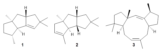
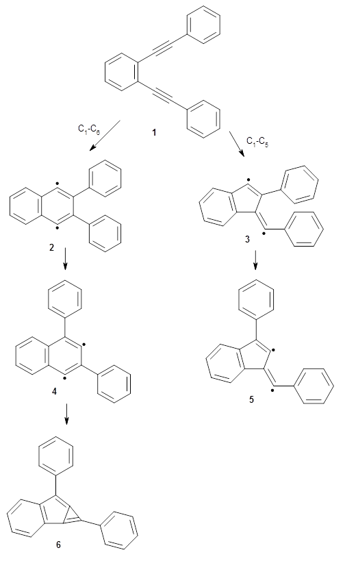
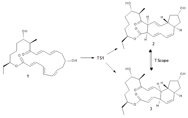
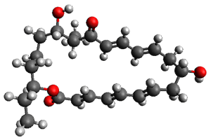
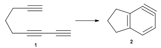
The first paper is by Skraba-Joiner, Johnson, and Agarwal.1 They discuss, among a number of interesting pericyclic reactions, the intramolecular Diels-Alder reaction of triyne 1 to give 2. They examined a concerted and stepwise pathway at (U)M05-2X/6-311+G(d,p) and find the concerted to be favored by 6.0 kcal mol-1. CCSD(T) using these geometries increases the difference to 8.2 kcal mol-1. The T1 diagnostic is fairly large for both the concerted and stepwise transition states, so they also performed CCSD(T)/CBS computations, which had much lower T1 values. The concerted TS remained favorable, but by only 2.7 kcal mol-1.

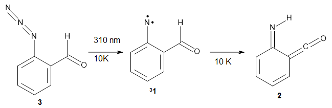
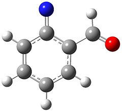
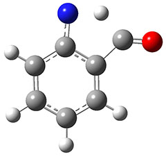
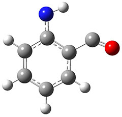
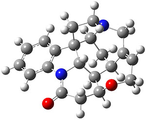
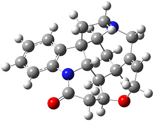
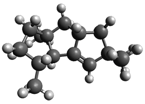
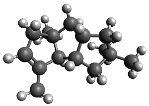
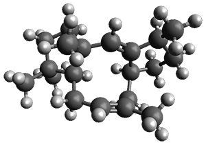
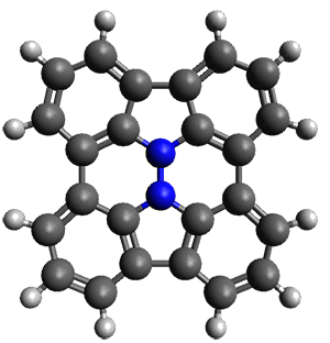
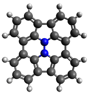
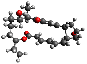
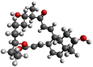
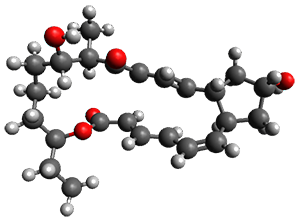
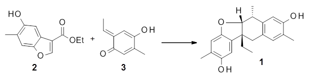
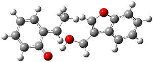
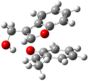
In the same special issue of the Journal of Organic Chemistry, Cramer, Hoye, and Kuwata examined a reaction closely related to what Johnson examined above.2 They looked at the reaction taking 3 into 4 via both experiments and computations. The M06-2x/6-311+G(d,p) geometries for the concerted and first TS along the stepwise path (with R1=R2=H) are shown in Figure 1. Evaluating the energies at SMD(o-dichlorobenzene)/B3LYP-D3BJ/6-311+G-(d,p)//M06-2X/6-311+G(d,p) find in this case (along with all of the other R1/R2 variants they examined) that the stepwise path has a lower barrier than the concerted path. In the case where R1=R2=H, the stepwise path is favored by 6.0 kcal mol-1. Additionally, these stepwise barriers are in reasonable agreement with the experimentally-derived barriers.

Concerted TS

Stepwise TS

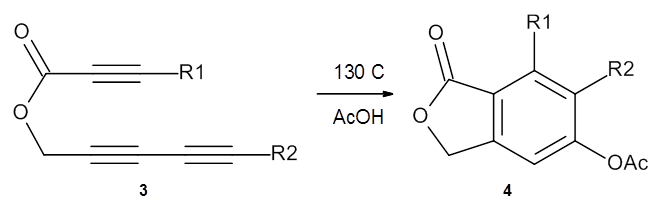
Figure 1. M06-2x/6-311+G(d,p) optimized geometries of the concerted and stepwise TSs for the reaction of 3H going to 4H.

It should be pointed out that the wavefunctions for the concerted TSs were all found to be unstable with regard to a restricted to unrestricted relaxation. Given this problem, they also performed a CASPT2 energy evaluation of the concerted and stepwise transition states for the case R1=R2=H. CASPT2 finds the stepwise barrier to be 3.7 kcal mol-1 lower than the concerted barrier.

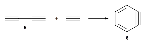
The last paper comes from the Houk lab, and examines the simplest set of intermolecular dehdro-Diels-Alder reactions.3 I will focus here on the most unsaturated analogue, the reaction of 1,3-butadiyne 5 with ethyne to give benzyne 6.

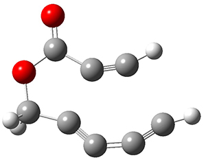
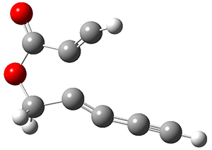
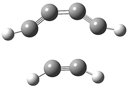
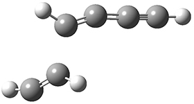
The concreted and stepwise transition states for this reaction (at (U)M06-2X/6-311+G(d,p)) are shown in Figure 2. The concerted barrier is 36.0 kcal moml-1 while the stepwise barrier is slightly lower: 35.2 kcal mol-1. The distortion energy for the concerted reaction is large (43.2 kcal mol-1) due mostly to angle changes in the diyne. Its interaction energy is -7.2 kcal mol-1, similar to the interaction energy in other similar Diels-Alder reactions. In contrast, the distortion energy for the stepwise pathway is 27.5 kcal mol-1, but the interaction energy is +7.7 kcal mol-1. These values are very similar to the distortion and interaction energy of the related (but less saturated DA reactions).

Concerted TS

Stepwise TS

Figure 2. (U)M06-2X/6-311+G(d,p) optimized concerted and stepwise TS for the reaction of 1,3-diyne with ethyne.

Molecular dynamics trajectories for both the concerted and stepwise paths reveal interesting differences. The concerted trajectories show an oscillatory behaviour of bending the angles at the C2 and C3 carbons prior to the TS, and then near synchronous formation of the new C-C bonds. The trajectories initiated at the stepwise TS show no systematic motion. Once the bond is formed, the biradical exhibits a long lifetime, on the order of picoseconds, much longer than the trajectory runs.

These three studies indicate the nature of the dehydro Diels-Alder reaction is very sensitive to reaction conditions, substituents, solvation, and all other manner of effects and will likely prove an area of interest for some time. It should keep a number of computational chemists busy for some time!

References

(1) Skraba-Joiner, S. L.; Johnson, R. P.; Agarwal, J. "Dehydropericyclic Reactions: Symmetry-Controlled Routes to Strained Reactive Intermediates," J. Org. Chem. 2015, 80, 11779-11787, DOI: 10.1021/acs.joc.5b01488.

(2) Marell, D. J.; Furan, L. R.; Woods, B. P.; Lei, X.; Bendelsmith, A. J.; Cramer, C. J.; Hoye, T. R.; Kuwata, K. T. "Mechanism of the Intramolecular Hexadehydro-Diels–Alder Reaction," J. Org. Chem. 2015, 80, 11744-11754, DOI: 10.1021/acs.joc.5b01356.

(3) Yu, P.; Yang, Z.; Liang, Y.; Hong, X.; Li, Y.; Houk, K. N. "Distortion-Controlled Reactivity and Molecular Dynamics of Dehydro-Diels–Alder Reactions," J. Am. Chem. Soc. 2016, 138, 8247-8252, DOI: 10.1021/jacs.6b04113.

InChIs

1: InChI=1S/C9H8/c1-3-5-7-9-8-6-4-2/h1-2H,5,7,9H2
InChIKey=IYZAZSVBWMMSLQ-UHFFFAOYSA-N

2: InChI=1S/C9H8/c1-2-5-9-7-3-6-8(9)4-1/h1,4H,3,6-7H2
InChIKey=PZJMTUKDGZUDBH-UHFFFAOYSA-N

3H: InChI=1S/C8H4O2/c1-3-5-6-7-10-8(9)4-2/h1-2H,7H2
InChIKey=MGXDIFXPYGGQLF-UHFFFAOYSA-N

4H: InChI=1S/C10H8O4/c1-6(11)14-8-2-3-9-7(4-8)5-13-10(9)12/h2-4H,5H2,1H3
InChIKey=GEFLHLNIKGXWCA-UHFFFAOYSA-N

5: InChI=1S/C4H2/c1-3-4-2/h1-2H
InChIKey=LLCSWKVOHICRDD-UHFFFAOYSA-N

6: InChI=1S/C6H4/c1-2-4-6-5-3-1/h1-4H
InChIKey=KLYCPFXDDDMZNQ-UHFFFAOYSA-N

« Previous PageNext Page »